Download
1 / 1
10 likes | 152 Views
Male femur. Width. Length. Sexual Dimorphism in Sea Spiders. College of Engineering and Natural Sciences. Kerry Quarry, Bonnie Bain, Fredric Govedich, Stephen Shuster. Department of Biological Sciences, Northern Arizona University, Flagstaff, AZ 86011 1. Additional Results. Abstract

E N D
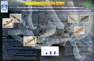
Male femur Width Length Sexual Dimorphism in Sea Spiders College of Engineering and Natural Sciences Kerry Quarry, Bonnie Bain, Fredric Govedich, Stephen Shuster Department of Biological Sciences, Northern Arizona University, Flagstaff, AZ 860111 Additional Results Abstract Pycnogonida (sea spiders) show varying degrees of sex role reversal. Males are smaller than females and possess specialized limbs (ovigers) for offspring care. Sex differences are thought to have arisen via sexual selection, making the Pycnogonida well suited for investigating this process, although sexual dimorphism in sea spiders is poorly known. We documented morphological Introduction Sea spiders (Arthropoda, Class Pycnogonida), are marine chelicerates found throughout the world in many different habitats including intertidal zones to the abyssal depths. Although spider-like in appearance, they are not true spiders, and are instead more closely related to the Acari, which includes ticks and mites. Sexes are separate in sea spiders and mature gonads in both males and females extend into the walking legs, usually to the distal end of the femur. Ammothea hilgendorfi (Cole, 1904) is a common west coast pycnogonid species, inhabiting intertidal zones from Tijuana, Mexico to San Francisco, California where it is usually found clinging on to the byssal threads of mussels or walking around on hydroid beds. Mature Female Immature Female Area differences in male and female walking legs in a common California pycnogonid, Ammothea hilgendorfi. Like most sea spiders, females in this species carry mature ova in the first 3 segments of their walking legs. The bulk of the ova lie within the femur. The male gonad is located in these same segments, but Both mature and immature female femurs had longest mid-femur widths and coxal end widths were longer than tibial end widths. occupies a much smaller volume than the female gonad. We measured femur length and 3 different femur widths (coxal end, midfemur, tibial end) for each specimen (n = 12) and calculated the area of each segment. Male and female femur lengths did not differ but females had longer midfemur/coxal end width and midfemur/tibial end width, and thus larger femur area compared to males. Immature and mature females had similar femur dimensions. Thus, while females with larger femurs may produce more offspring than females lacking such a modification, we cannot eliminate the hypothesis that selection has not also favored narrower femurs in males. Egg size and egg number did not show a significant relationship to femur size. Results Methods • 209 specimens were collected over a period of one year near Mission Bay, San Diego, CA, USA. Sex ratio was determined to be 50:50 based on the entire collection. • All specimens from one collecting trip (n = 75) were used to prepare whole mounts for further study. One walking leg per individual was made into a whole mount using standard techniques. Of the 75 specimens, four were males and 71 were females. Conclusions • Mature females and males are sexually dimorphic. • Femur area is larger in females than in males due to greater female femur width than males; femur lengths were not significantly different between the sexes. • Females had longer midfemur/coxal end width and midfemur/tibial end widths compared with males. Male femurs are more tapered at each end than female ones. • Both immature and mature females had similar femur measurements, indicating female femur shape is independent of sexual maturity. Both mature and immature females had relatively long midfemur widths and a longer coxal end width than tibial end width. • Egg size and egg number did not have a significant relationship with femur size, demonstrating that increased femur size does not increase fecundity. Femur Measurements • Mature and immature females were separated based on the number and size of eggs within the femur. Four mature and four immature females were randomly selected for comparison to the four males. • Photographs were taken with Motic Educator. • Femur length (coxal end-tibial end) and width (midfemur) were measured with ImageJ • Area was determined by length x width. • For 4 males, 4 mature, and 4 immature females, additional width measurements were taken (coxal end width; tibial end width) • Female femurs contained ova in 2 size classes (large, small) • Egg circumference was measured in 21 females; all visible femur eggs were measured (max. = 21 eggs per femur) and radius was derived from circumference using: circumference = 2*pi*radius References Cole, L. J. 1904. Pycnogonida of the West Coast of North America. Harriman Alaska Expedition, 10: 249-330. Bain, B., 1991. Some observations on biology and feeding behavior in two southern California pycnogonids. Bijdragen tot de Dierkunde, 61 (1) 63-64.