Download

1 / 37
400 likes | 578 Views
MRI IN CHILDREN 2014. DR JAYAPRAKASH KP,ASST PROF,ICH,MCH,KOTTAYAM. LEARNING OBJECTIVES. List the advantages of MRI over other existing modalilities of imaging Compare type of images in MRI and their advantages List 4 situations where T1 image is useful

E N D
MRI IN CHILDREN 2014 DR JAYAPRAKASH KP,ASST PROF,ICH,MCH,KOTTAYAM
LEARNING OBJECTIVES • List the advantages of MRI over other existing modalilities of imaging • Compare type of images in MRI and their advantages • List 4 situations where T1 image is useful • List 4 situations where T2 images are beneficial • identify 2 conditions where FLAIR has definite advantage • List 2 conditions where DWI has diagnostic value • Identify 2 situations where MR spectroscopy is applied • Compare the advantage of functional MRI with conventional MRI
Magnetic resonance imaging (MRI) is based on the absorption and emission of radiofrequency energy by hydrogen protons whose spin is influenced by changing magnetic fields (0.3 to 1.5 T). Unlike computed tomography (CT), there is no radiation exposure
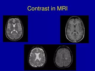
T1-weighted images cause fat (eg, myelin in white matter) to appear bright and water (eg,cerebrospinalfluid [CSF] or edema) to appear dark on this sequence. The gray-white interfaces of the brain are well depicted on these sequences, especially if with the images are thinly sliced. • T2-weighted images cause water (eg, CSF and edema) to appear bright and fat to appear dark. The MRI-based intravenous contrast agents (eg, gadolinium) are frequently used in T1-weighted images (Fig 1A and B) to make serum appear bright
T1-weighted image at the level of midbrain. A. The cerebrospinal fluid (CSF) appears dark. B. The CSF appears bright.Notethe gray and white matter differentiation in the 2 images. The anatomical structures identified here are the caudate nucleus (i), lenticular nuclei (ii), thalamus (iii), frontal horn of lateral ventricle (iv), and atria of the lateral ventricle (v).
Case 1 • A 7-year-old girl with a history of migraine headaches presented with a head tilt to the left and worsening headaches. Optic disc edema was found on ophthalmoscopy. • A brain MRI was performed, which revealed a large heterogeneous intermediate signal mass that filled and obstructed the fourth ventricle on the axial T2-weighted images at the level of the fourth ventricle . She underwent surgical resection, and the histologic diagnosis was medulloblastoma. • Apart from sparing the effects of radiation, MRI is superior to CT in delineating tumor extent, spread, mass effect, vascularity, necrosis, and edema. T2-weighted sequences are sensitive for the detection of tumor and edema.
Axial T2-weighted image at the level of the fourth ventricle shows a large heterogeneous intermediate signal mass (blue arrow) that fills and obstructs the fourth ventricle.
Case 2 • A 6-year-old girl presented with swelling of her right eye. She had a history of sinusitis. Her right eye was swollenand erythematous, she had difficulty opening her right eye, and she had double vision with all gazes. • Clinical findings included proptosis, ptosis, restriction of ocular motility, ocular pain, and chemosis. Laboratory tests revealed neutrophilicleukocytosis and elevated C-reactive protein. • Head MRI was performed. Coronal postcontrast T1-weighted imaging revealed inflammation in the rightorbitwith diffuse stranding in orbital fat, enhancing myositis of the inferior and medial rectus muscles
Coronal postcontrast T1-weighted image shows inflammation in the right orbit with diffuse stranding in orbital fat, enhancing myositis of the inferior and medial rectus muscles (solid yeCllowarrow indicates the right inferior rectus; solid blue yellow indicates the right medial rectus muscle).
Orbital cellulitis is an infection of the soft tissue posterior to the orbital septum, whereas preseptalcellulitis affects anterior to the orbital septum. The former is distinguished from the latter by the presence of proptosis,chemosis, ophthalmoplegia, or decreased visual acuity. • MRI is superior to CT when there is suspicion of intracranial extension, optic nerve involvement, and cavernous sinus thrombosis because MRI is better for discerning soft tissue disease. • Gadolinium is a paramagnetic contrast agent that prolongs the spin of water protons, resulting in postcontrast enhancement of areas of inflammation on T1- weighted imaging. The most sensitive technique for demonstrating orbital infection is post gadolinium, fat suppressed T1-weighted imaging.
Case 3 • A 16-year-old girl presented to the emergency department with a chief symptom of “vision loss in right eye.” She denied eye pain, diplopia, photophobia, or headacheHer medical history was unremarkable. Visual acuity was normal in the left eye (20/20); however, it was decreased • in the right eye (20/200). Her right pupil constricted in response to consensual but not to direct light (ie, deafferented pupil). Bilateral fundi appeared normal. MRI of the brain revealed a normal-appearing left optic nerve. • The left optic nerve was round with distinct borders and was appropriately surrounded by CSF as demonstrated by T2-weighted fluid-attenuated inversion recovery (FLAIR) imaging. The right optic nerve, however, had poorly defined borders, suggesting an inflammatory process. The inflamed right optic nerve also enhanced with gadolinium administration
The left optic nerve (solid yellow arrow) is round with distinct borders and is appropriately surrounded by cerebrospinal fluid as demonstrated by T2-weighted imaging. The right optic nerve (solid blue arrow), however, has poorly defined borders, suggesting an inflammatory process
Given the patient’s visual disturbance and the evidence of optic nerve inflammation on MRI, optic neuritis was suggested as a iagnosis. Other MRIs obtained revealeddemyelinationin the pons and cerebellum. Thepresenceof CSF oligoclonal bands supported multiplesclerosisas the diagnosis. • FLAIR is an extremely useful technique in brain imaging.Likeconventional T2-weighted imaging, edema appearsbright, but this technique nulls (or makes dark)CSF Signal. • FLAIR is a sensitive technique for displaying demyelination within the brain, thus clearly revealing lesions in proximity to CSF, such as periventricular plaques in multiple sclerosis. The technique is accomplished via a relatively long inversion time to allow the longitudinal
The technique is accomplished via a relatively long inversion time to allow the longitudinalmagnetization of CSF to return to the null point preceding the conventional spin echo imaging. It also has a tremendous role in early detection of cortical gray matterinfarcts. • The cortical gray matter is vulnerable to ischemiabecauseof its high metabolic activity. However, corticalgraymatter immediately djacentto CSF within the sulci makes infarction hard to delineate when this area undergoesconventionalimaging equencesthat emphasizefluidsignal. • FLAIR suppresses the CSF signal and makesthecortical or periventricular area more conspicuous
The left optic nerve (solid yellow arrow) is round with distinct borders and is appropriately surrounded by cerebrospinal fluid as demonstrated by T2-weighted imaging. The right optic nerve (solid blue arrow), however, has poorly defined borders, suggesting an inflammatory process
Case 4 • A 3-month-old boy was admitted to the pediatric intensive care unit for bilateral subdural hematomas (SDHs)and concern for intentional trauma. Per his parents, hehademesis for 24 hours and had been unable to keep formuladown. He also had 2 episodes of arm stiffening and breath holding followed by agitation and crying. Neurologicexaminationrevealed an enlarged head circumference,dilatedscalp veins, and a bulging anterior fontanel. • He had brisk tendon reflexes. Brain MRI FLAIR imagesrevealedlocalizing tissue loss in the right parietal regionfroman older injury and 2 SDHs of different densities,withthe more acute-appearing SDH on the left andthemore subacute SDH on the right
The presence of SDHs of varying ages and of skull fractures, which are depressed or multiple or diastatic or involvemultipleor nonparietal bones, is a key neuroimaging finding that is consistent with the diagnosis of intentionaltrauma. CT is the modality of choice to detect acute hemorrhagesandskull fractures. • MRI is superior to CT in detecting extra-axial hemorrhages, diffuse axonal injury, andearlyrecognition and prognostication of parenchymal injury.SDHsover the falx, posterior fossa, and tentoriumare more characteristic of intentional trauma. • (1) T2-weighted gradient echo MRI enhances the sensitivity for recognizing acute bleeds and Old shear bleeds. Diffusion-weighted imaging (DWI) is also helpful for revealing early and progressive edema. Of note, bleeding diathesis, birth injury, inborn errors of metabolism (such as glutaric academia type 1 and Menkes disease), lymphohistiocytosis, infection,andunintentional trauma can have similar appearance with DWI.
Fluid-attenuated inversion recovery image localizing tissue loss in right parietal region from an older injury and 2 different densities of subdural hematoma (more acute appearing on the left and subacute on the right).
Case 5 • A 4-year-old previously healthy girl was admitted for leftsided weakness and increased somnolence. A week beforepresentation, she had severe gastroenteritis. Her oral intakewasdrastically reduced, and she had presented to a local emergency department 2 days earlier with persistent vomiting. On the day of her presentation, she was unabletowalk. Her vital signs were stable. • On examination, shewasdrowsy. She had left-sided upper motor neuron facial weakness. Her muscle strength in the left upper andlowerextremities was 0/5 and 1/5, respectively, and she had a positive Babinski sign. The rest of her examinationfindingswere unremarkable. Her head CT was unremarkable.MRIand magnetic resonance venography (MRV) of the brain demonstrated a large filling defect,occludingand occupying the superior sagittal sinus andmedialportion of both transverse sinuses. • A conventionalheadMRI revealed venous infarctions in the left parietal and frontal areas without any hemorrhage.Hercoagulation profile was normal. She was treated with intravenous heparin and fluids. Her weakness improvedgradually, and subsequent MRV revealed restoration of normal contrast enhanced filling of sagittal and bothtransverse sinuses
MRVis the optimal noninvasive technique of delineating cerebral venous anatomy. Time-of-flight, phase-contrastangiography, and contrast-enhanced MRV are themeanscommonly used to evaluate the cerebral venousstructures, commonly the superior sagittal, ransverse, sigmoid, and straight sinuses. • MRI andMRV are importantfordemonstrating venous occlusion and its consequences,infarction, and edema. Of note, a thrombus in its subacute phase can be recognizable as a high signal on a T1-weighted scan, thereby making MRV unnecessary to perform for a suspected clot sigmoid, and straight sinuses. • MRI andMRV are important for demonstrating venous occlusion and its consequences, infarction, and edema. Of note, a thrombus in its subacute phase can be recognizable as a high signal on a T1-weighted scan, thereby making MRV unnecessary to perform for a suspected clot.
A. Magnetic resonance Venography (MRV) revealslargefilling defect that occludes and occupies the superior sagittal sinus (broken blue arrow) and medial portion of both transverse sinuses (broken yellow arrows). B. On the right, coronal MRV with contrast reveals restoration of normal contrast enhanced filling of sagittal (solid blue arrow) and both transverse sinuses (solid yellow arrows
Case 6 • A term infant developed right-sided clonic seizures on her second day of life. The pregnancy was well supervised, and her delivery was uneventful. Her physical examination findings were normal on the first day of life. Maternaldrugscreen result was negative. Metabolic profile, bloodcellcount, transfontanel ultrasonography, and CSF studyresultswere normal. A head MRI was performed. • Coronal T2 revealed left-sided loss of gray white differentiationandswelling in the region of the posterior cerebral artery with obscuring of the ventricular surface (ventricular effacement). The hyperintense signal in the posterior cerebral artery region on DWI and the hypointense signal in the same distribution on apparent diffusion coefficient imagingconfirmed the presence of restricted diffusion characteristics of an evolving acute infarction
By disrupting the cellular metabolism and Naþ/Kþadenosine triphosphatase pump, ischemia results in lossoftransmembrane ionic gradients, thereby restricting diffusionofwater through cellular membranes. • DWI was performed by adding 2 strong diffusion-sensitizing magneticfieldgradient pulses. DWI depicts recently infarcted brain as a very bright signal. The actual diffusion in the tissueisdecreased as seen on apparent diffusion coefficient maps. This period of restricted diffusion can last 5 to 10days (but sometimes less) in the pediatric population. • DWI can detect ischemic stroke withinminutes, in contrasttoconventional MRI, which may take hours for diagnosis. DWI is invaluable in detecting lesions not usually identifiable with conventional MRI and can distinguish new and old strokes and acute andchronicones. Because abscesses, parenchymal contusions, and cysts canalsodemonstrate restricted diffusion, DWI also helps detect brain abscesses and cystic tumors.
Coronal T2-weighted image shows loss of gray white differentiation and swelling in the posterior cerebral artery territory on the left side with ventricular effacement secondary to swelling.
A. Hyperintense signal in posterior cerebral artery territory on diffusion-weighted imaging. B. Apparent diffusion coefficient image has marked hypointense signal in the same distribution, confirming restricted diffusion characteristics of acute evolving infarct.
Other MRI Modalities • Magnetic resonance spectroscopy monitors biochemical changes inbraintumors, head trauma, stroke,epilepsy, metabolic disorders, andinfections. • The metabolites predominantlymeasuredareN-acetylaspartate (NAA), creatine, choline, and myoinositol.NAA, an amino acid foundexclusivelyin neurons, is regarded as a nonspecific marker of neuronal viability. NAA levels are decreased in conditions such as infarction and neuronal inflammation. Lactate is absent in normal brain tissue, and its presence is indicative of anaerobic glycolysis at the cellular level. rs(with predominant anaerobElevated lactate levels are associated with ischemia or neurometabolicdisordeic
A 16-year-old with a 1-week history of “bumping into things.” Contrastenhanced magnetic resonance imaging (MRI) shows an enhancing lesion in the occipital lobe. MRI spectroscopy reveals decreased N-acetylaspartate peak, maintained cholinecreatine ratio, and the presence of a large lactate peak. These findings are indicative of an ischemic stroke.
with predominant anaerobic glycolysis Elevated lactate levels are associated with ischemia or neurometabolicdisorder). An elevated choline-creatine ratio is suggestive of malignancy. • Choline is involved in the synthesis of phospholipid cell membranes; in aggressive neoplasms,cholineis elevated because of rapid cell turnover. • Creatine,aprecursor of adenosine triphosphate, gives a measureofbrain energy stores. In high-grade brain tumors,dueto increased metabolic activity, creatine is depleted. shows magnetic resonance spectroscopy image from a 16-year-old girl with ischemic stroke in the occipitalarea. There is a large lactate peak, decreased NAApeak, and maintained choline-creatine ratio. These findings are characteristic of an ischemic stroke.
FunctionalMRI (fMRI) reveals brain activity by changing magnetization between oxygen-rich and oxygen-poor blood. fMRI is useful for presurgical mapping, epilepsy,stroke, trauma, tumors, language lateralization, and neurobehavioral • disorders, such as attention-deficit/hyperactivitydisorder(ADHD) and autism. The diagnosis and management of ADHD can be a challenge because diagnosisrelieson parents’ and teachers’ reporting of behavioral • symptoms that might be biased and subjective. fMRIhasshown that in patients with ADHD there is reducedactivationof striatum and frontal lobes during tasks that warrant attention. Thus, in the future, fMRI could beusefulto diagnose, prognosticate, and treat patients withADHD.
Children with ventriculoperitoneal shunts for hydrocephalus require subsequent follow-up CT to evaluate shunt integrity, assess ventricle anatomy, and rule outcomplications. However, multiple lifetime exposure oionizingradiation is associated with potential risk for carcinogenesis. • Single-shot fast-spin echo (quick-brain)MRI can be used in children with shunt-treated hydrocephalus for workup and follow-up in lieu of CT to circumvent radiation exposure. Because of its ability to generate required data with a single excitation impulse, • single-shot fast-spin echo is fast and does not require sedation.Multiplanarimages are obtained in less than a second,reducingthe motion artifacts.
MRI vs a Good History and Physical Examination • Although MRI can be significantly informative, a good history and clinical examination are sufficient for diagnosing and managing many clinical situations, such as primary headache (chronic or recurrent headache, including migraine without permanent neurologic signs or signs of increased intracranialpressure), simple syncope, simple febrileseizures, and benign positional vertigo
Summary • • On the basis of strong recommendation, magnetic resonance imaging (MRI) and computed tomography(CT) are complementary diagnostic tools with mutually distinct advantages and pitfalls. MRI ispreferredto CT in posterior fossa disease, white matterdisease, temporal lobe epilepsy, and vascular diseases. • • On the basis of strong recommendation, diffusionweightedimagingis superior to noncontrast CT fordiagnosisof acute ischemic stroke in patients who present within 12 hours of symptoms. (5) • • On the basis of strong recommendation, fluidattenuatedinversionrecovery MRI is a sensitive technique for displaying demyelination within thebrain, especially in lesions near the cerebrospinal fluid,suchas periventricular plaques in multiple sclerosis. In clinically isolated syndrome, 3 or more white matterlesionson T2-weighted MRI is highly predictive of future development of clinically definite multiplesclerosis. (6) • • There is insufficient evidence to support or refute neuroimaging in a child presenting with status epilepticus
1. An infant in the newborn nursery has just experienced 3 focal seizures of her right arm, yet she appears well and is afebrile. You suspect a neonatal stroke. Among the following, the MOST appropriate brain imaging to • establish this diagnosis is: • A. Computed tomography. • B. Magnetic resonance angiography. • C. Magnetic resonance imaging with diffusion-weighted imaging. • D. Magnetic resonance spectroscopy. • E. Single-shot fast-spin echo magnetic resonance imaging
2. You are counseling the parents of a toddler with shunted hydrocephalus. His family is concerned about the • long-term effects of radiation exposure used to image the brain for evidence of shunt malfunction. Among the • following, you are MOST likely to say: • A. Computed tomography has a limited radiation exposure, far less than therapeutic radiation, and the couple • should not be concerned. • B. Head ultrasonography should always be used as a screening tool before considering computed tomography. • C. Magnetic resonance imaging with diffusion-weighted imaging will be useful to distinguish acute from • subacute shunt failure. • D. Magnetic resonance imaging with fluid-attenuated inversion recovery (FLAIR) differentiates • cerebrospinal fluid as dark from other water, which appears bright, and FLAIR is an optimal way to • diagnose shunt failure. • E. Single-shot fast-spin echo magnetic resonance imaging is a promising technique to obtain imaging • in just seconds without sedation and is optimal to evaluate shunt integrity and assess ventricular • anatomy.
3. A 6-year-old presents with progressive occipital headaches, nausea, and vomiting for 2 weeks. Head • computed tomography in the emergency department reveals a possible lesion in the posterior fossa. You • now request brain magnetic resonance imaging with gadolinium contrast because this contrast will help • highlight: • A. Arachnoid cyst. • B. Cerebral dysgenesis. • C. Chiari malformation. • D. Hydrocephalus. • E. Tumor.
4. A 15-year-old boy presents to the emergency department with severe headache after striking his head without a helmet while skateboarding. You decide to perform imaging of his head. Among • the following, the MOST appropriate modality to detect a skull fracture or acute parenchymal • hemorrhage is: • A. Computed tomography. • B. Magnetic resonance imaging with diffusion-weighted imaging. • C. Magnetic resonance spectroscopy. • D. Single-shot fast-spin echo magnetic resonance imaging. • E. Ultrasonography.
5. A 16-year-old girl was found to have a right parietal mass with computed tomography. She is scheduled to undergo magnetic resonance imaging, and the radiologist suggests adding spectroscopy to determine the nature of the mass. Among the following, the finding MOST suggestive for tumor on spectroscopy is: • A. Decreased lactate. • B. Decreased phospholipid. • C. Elevated choline-creatine ratio. • D. Elevated creatine. • E. Elevated N-acetyl aspartate