Download
1 / 38
410 likes | 735 Views
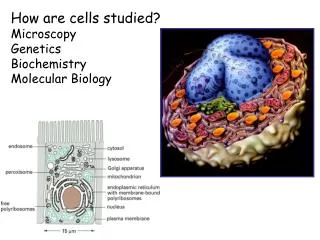
How are cells studied? Microscopy Genetics Biochemistry Molecular Biology. Light microscopy allows examination of cell morphology. Cells are highly diverse. Cell shape is determined by a cell wall, or by the cytoskeleton. A protozoan (Didinium) eating another. Bars 10 µm. dinoflagellate.

E N D
How are cells studied? Microscopy Genetics Biochemistry Molecular Biology
Cells are highly diverse Cell shape is determined by a cell wall, or by the cytoskeleton A protozoan (Didinium) eating another Bars 10 µm dinoflagellate euglenoid B cells T cells ciliates amoeba heliozoan
Cell compartmentalization is achieved by the use of membranes, which are composed of phospholipid bilayers. Membranes make life on Earth possible, but they also present a great problem, as they impose barriers to diffusion and intracellular transport
Biological membranes- (e.g. the plasma membrane)- 3. function The membrane encapsulates cellular components and maintains an equal solute concentration between the inside and the outside of the cell. A biological membranes’ main function is to segregate chemicals. Outside 35-50 Å Inside Membranes impose barriers to diffusion 2. morphology 1. fluidity
Chemical nature of phospholipids- Phosphatidylcholine
Phospholipids are amphiphilic molecules- Hydrophilic and hydrophobic molecules interact differently with water …
Lipids assemble spontaneously into sheets, liposomes and micelles- Lipids self-associate without covalent bonding; their tails cooperate to exclude water A lipid’s chemistry determines its geometric shape (e.g. cones, cylinder, etc.)
Different kinds of phospholipids- * Note their asymmetric distribution in the two membrane leaflets
General function of biological membranes as semi permeable barriers
Membranes function as selective chemical barriers- Membrane permeability
The water channel- Discovery of these water channels led to a Nobel Prize in Chemistry in 1993 to Dr. Peter Agre
Intracellular membranes serve as physical barriers that allow compartmentalization- Membranes everywhere…
The fluid mosaic model of membrane composition & Topology of membrane associated proteins
Proteins are embedded on membranes via hydrophobic surfaces- Structure of an alpha helixusually 20 amino acids long Hydropathy plot Structure of a beta barrel Hydrophobic tails Transmembrane domains Glycolipid anchor Fatty acid anchor
Biol110L-Cell Biology Lab-Spring 2011 Module #1: Cell morphology and organelle compartmentalization B. Membrane structure and function C. Cellular fractionation and protein topology
A. Cell morphology and organelle compartmentalization Budding yeast (Saccharomyces cerevisiae) is a model eukaryotic cell Our experimental organism of choice
Lipophilic dyes can be used to visualize membranes- DiOC6 (high concentration) Mitochondria, ER, etc DiOC6 (low concentration) mitochondria
Fluorescence microscopy using GFP Green fluorescent protein (GFP) Useful when you want to find out the location of a particular protein in cells, to a radius of ~200 nm of its locale You need to make a gene fusion between the genes encoding GFP and your protein of interest Cells are not fixed prior to visualization of cells under the microscope; therefore, the technique is used when you want to visualize a protein (a fusion protein) in ‘real time’
A collection of yeast strains, each carrying a single GFP tagged protein… Access the database at- http://yeastgfp.yeastgenome.org/ Access the S. cerevisiae database at- http://www.yeastgenome.org/ for information on each protein Cell periphery YLR413W (n/a) YLR332W (Mid2p) YEL063 (Can1p) YMR058W (Fet3p)
Mitochondria YER080W (Fmp29p) YOR356W (n/a) YGL068W (n/a) Nuclear periphery YML031W (Ndc1) YML075C (Hmg1) YOR046C (Dbp5)
Spindle pole YGL061C (Duo1p) YDR320C (Dad4p) Nucleoplasm YER156c YGL097w (Prp20p)
Nucleolus Ygl078c (Dbp3p) Yol077c (Brx1p) Cis-Golgi Yfr051c (Ret2p) Ynl287 (Sec21p)
Vacuole Ydl185w (Tfp1p) Yor332w (Vma4p) Cytosol Ymr235c (Rna1p) Yll024c (Ssa2p)
B. Membrane structure and function To expose the yeast plasma membrane for analysis and to weaken the cells in preparation for cell fractionation, we must first remove the tough yeast cell wall
Yeast cell wall composition The cell wall can be removed with lyticase: a beta 1,3 glucanase (originally obtained from the gut of snails)
If you want to fractionate cells to isolate an organelle or to determine the cellular distribution* of a protein, use differential velocity sedimentation Differential velocity sedimentation resolves particles based on size (3,000 x g) Low speed pellet LSP (15,000 x g) Medium speed pellet MSP (100,000 x g) High speed pellet HSP ---> ribosomes, large macromolecules Low speed supernatant LSS Medium speed supernatant MSS High speed supernatant HSS ---> small soluble proteins & molecules
Different types of membrane proteins- Term used for each protein in this intracellular membrane compartment: #1: lumenal soluble protein #2: lumenal peripheral membrane protein #3: transmembrane or integral membrane protein (single pass or multi-pass) #4: cytosolic monotopic-integral membrane protein #5: cytosolic peripheral membrane protein #6: cytosolic calcium-dependent peripheral membrane protein #7: cytosolic peripheral membrane protein #8: cytosolic lipid-anchored peripheral membrane protein
Detergents solubilize membranes by dispersing their phospholipids Membrane solubilization with Triton X-100 + + Detergents Triton X-100 (a non-ionic detergent) dissolves membranes and solubilizes membrane proteins without affecting their structure/ function. SDS (an ionic detergent) dissolves membranes and denatures protein structure.
Characterization of protein topology on biomembranes- Subject membranes to centrifugation, which separates soluble (S) from insoluble (or membrane bound or membrane enclosed) material (P).
Example: Samples from chromatographic column fractions are analyzed during purification of a protein Analysis of the protein composition of a solution by SDS-PAGE (polyacrylamide gel electrophoresis)- Used to look at the protein composition of a biological sample. Stain with Coomassie for visualization in the gel Perform western blot to identify one protein amidst many
If you want to visualize a single known protein within a collection of proteins…use Western blotting with specific antibodies- Transfer proteins from an SDS-PAGE gel to nitrocellulose or PVDF membranes (using electrophoresis), then blot as shown below….
Cell osmolarity- solute concentration macromolecules organic molecules ions
Cellular mechanisms for dealing with osmolarity issues- Cell wall and turgor pressure in plants Active ion pumps Water extrusion