Download
1 / 56
560 likes | 728 Views
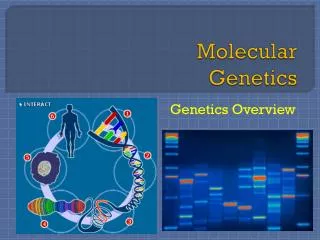
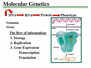
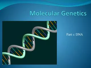
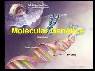
Section A Molecular Genetics. Organism. System. Organ. Tissue. Cell. Cellular Organelle. Molecule (DNA, RNA, protein, etc). Content. A1 DNA structure A2 RNA structure A3 Transfer RNA (tRNA) A4 Ribosomal RNA (rRNA) A5 Messenger RNA (mRNA). A6 DNA replication A7 Gene transcription

E N D
Organism System Organ Tissue Cell Cellular Organelle Molecule (DNA, RNA, protein, etc)
Content • A1 DNA structure • A2 RNA structure • A3 Transfer RNA (tRNA) • A4 Ribosomal RNA (rRNA) • A5 Messenger RNA (mRNA)
A6 DNA replication • A7 Gene transcription • A8 The genetic code • A9 Translation • A10 Regulation of gene expression in prokaryotes • A11 Regulation of gene expression in eukaryotes
A1 DNA STRUCTURE • Nucleotides['nu:kli:ə,taɪd, 'nju:-] • DNA polynucleotides • The double helix • Complementary base pairing • RNA structure
观点:DNA序列以此为基本单位组成,无变化, 不可能是遗传物质
What is the genetic material that passing on to make new offspring ?
Griffith’s Experiment: Pneuococcus transformation
“DNA isthe true genetic materials”--- Confirmation by the infection experiment of bacteria phage
RNA also can be genetic material (virus). • Electron micrograph of tobacco mosaic virus. Magnification 37,428. • Reconstitution experiment of Fraenkel-Conrat and Singer. The nucleic acid (RNA), not the protein component of the virus, controls inheritance.
DNA has many features that make it useful as genetic material: (1) It can store plenty of genetic information in its base sequence; (2) It can transfer this information to RNA and protein by transcription and translation; (3) It has great physical and chemical stability; (4) DNA can self replicate, passing genetic information to progeny cells/offsprings; (5) It can bring mutational alterations causing variation, at the same time, occurring without loss of parental information.
the structure of DNA and RNA
1. Nucleotides • The ability of DNA to carry genetic information closely related to its structure • The DNA molecule consists of a long chain of monomers called nucleotides, thus, DNA is called a polynucleotide. • The components of each nucleotide: a sugar, a base and a phosphate group
Bases(Fig. 1 a) adenine ['ædəni:n](A), guanine ['ɡwɑ:ni:n](G)…..Purines thymine ['θaimi:n](T), cytosine [‘saitəsi:n](C)......Pyrimidines [,pai'rimidi:n] • Sugar: 2′-deoxyribose (Fig. 1b) • Phosphate group
T U C
Nucleoside ['nju:kliəsaid] : a sugar plus a base (Fig. 2) • Covalent bond between • 1′ carbon of the sugar and • a nitrogen ['naitrədʒən] at position 9 of the purines or • position 1 of the pyrimidines
Nucleotides: one to three phosphate groups (PO4) plus a nucleoside (Fig 2) • PO4 attached to the 5’ carbon of the sugar • Four nucleotide triphosphates: • 2’-deoxyadenosine 5’-triphosphate (dATP or A), • 2’-deoxythymidine 5’-triphosphate (dTTP or T), • 2’-deoxycytosine 5’-triphosphate (dCTP or C), • 2’-deoxyguanosine 5’-triphosphate (dGTP or G)
2. DNA polynucleotides • Nucleotide triphosphates are joined together to give polynucleotides • 3’-5’ phosphodiester bond (C-O-P) (Fig. 3a) • Primary structure of DNA molecule (Fig. 3b)
Figure 3. 3’ to 5’ phosphodiester bond (a) and DNA primary structure (b)
Free 5’-end (5’ triphosphate) and free 3’-end(3’ hydroxyl group): polarity • The sequence of the bases in the DNA polynucleotide encodes the genetic information, which is written in the 5’ →3’ direction. • No apparent limit to the number of nucleotides and on the sequence of them
1940s-1950s • Experiments provide evidence • DNA is the genetic material,but • Why could DNA be used as genetic material? • How does it work? • Understand its structure Structure is the basis of function…… Many scientists were attracted to reveal the structure of DNA, including Watson and Crick
Double-Helix DNA model—Evidence 1: Purine and Pyrimidine have equal molar weight.
Double-Helix DNA model—Evidence 2: • The X-ray diffraction pictures of DNA taken by Franklin and Wilkins provided direct evidence.
In 1953, Watsonand Crick, working in Cambridge, England, established the three-dimensional structure of DNA • The double helix structure of DNA
James D. Watson (1928– ) (Cold Spring Harbor Laboratory Research Library Archives. Margot Bennet, photographer.) Francis Crick (1916–2004) (Reproduced by permission of Herb Weitman, Washington University, St. Louis, Missouri.) Rosalind E. Franklin (1920–1958)
Francis Crick James Watson
The DNA double helix model • DNA exists as two polynucleotide chains wrapped around each other to form the double helix; • The sugar-phosphate part of the molecule forms a spindle ['spɪndl] or backbone which is on the outside of the helix; • The bases, which are flat molecules, face inwards towards the center of the helix and
are stacked on top of each other like a pile of plates. • The double helix executes [,eksi'kju:ts] a turn every 10 base pairs. The pitch of the helix is 34 Å so the spacing between bases is 3.4 Å. The diameter of the helix is 20 Å. • The two polynucleotide chains are antiparallel. Major and minor grooves exist. • The double helix is right-handed.
Variant forms of DNA (Fig. 6) • Under different conditions the crystals of the molecule display various forms. • B form and A form both are right-handed. • Left-handedhelix: Z form
4. Complementary base pairing • The bases of the two polynucleotide chains interact with each other in the form of hydrogen bonds to stabilize the interaction. • Two bonds between A and T and three bonds between G and C (Fig. 7). • The way in which the bases form pairs between the two DNA strands is known as complementary base pairing and is of fundamental importance.
The sequences of the two strands are related to each other and are said to be complementary with the sequence of one strand predicting and determining the sequences of the other. The basis of DNA replication. Essential for expression of genetic information: from DNA to mRNA (transcription)
Hydrogen bond can be disrupted by heat and some chemicals, causing the separation of the double helix into two strands: denaturation • In cells, for replication and transcription, the strands of double helix is transiently and dynamically separated.
5. RNA (primary) structure • The component of RNA is different from that of DNA in that ribose replaces 2-deoxyribose and the base uracilreplaces thymidine. • RNA molecules normally exist as a single polynucleotide strand, while short double-stranded regions formed pairing between bases within the same RNA strand (Fig.8).