Download
1 / 82
950 likes | 1.68k Views
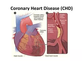
Coronary Heart Disease (CHD). Leading cause of death in U.S. Narrowing coronary arteries Atherosclerosis. Angina Pectoris - Pathophysiology. Obstructed coronary artery Increased myocardial oxygen demand Lactic acid release Leads to pain Three types Stable Unstable

E N D
Coronary Heart Disease (CHD) • Leading cause of death in U.S. • Narrowing coronary arteries • Atherosclerosis
Angina Pectoris - Pathophysiology • Obstructed coronary artery • Increased myocardial oxygen demand • Lactic acid release • Leads to pain • Three types • Stable • Unstable • Prinzmetal’s: is a syndrome typically consisting of angina (cardiac chest pain) at rest that occurs in cycles. It is caused by vasospasm, a narrowing of the coronary arteries caused by contraction of the smooth muscle tissue in the vessel walls rather than directly by atherosclerosis
Angina Pectoris - Manifestations • Chest pain • Radiates • Onset with exercise, etc. • Relieved by rest, nitroglycerin (NTG) • SOB, pallor, fear
Acute Coronary Syndrome • Condition that includes: • Unstable angina • Acute myocardial ischemia with or without muscle damage • Associated with coronary artery stenosis and atherosclerotic plaque
Acute Myocardial Infarction (AMI) • Pathophysiology • Occluded coronary artery stops blood flow to part of cardiac muscle • Cellular death • Tissue necrosis • Description—heart area affected • Classification
AMI Manifestations • Chest pain • Radiates to shoulder, neck, jaw, arms • Lasts longer than 15–20 minutes • Not relieved with NTG • Sense of impending doom • SOB • Diaphoresis • Nausea and vomiting
AMI Manifestations (continued) • Manifestations in women and elderly • May be atypical • Upper abdominal pain • No chest pain but other symptoms
AMI Complications • Related to size and location of infarct • Dysrhythmias • Pump failure • Cardiogenic shock • Pericarditis
Cardiac Dysrhythmias • Pathophysiology • Due to altered formation of impulses or altered conduction of the impulse through the heart • Ectopic beats • Heart block • Reentry phenomenon • Classified to the site of impulse formation or the site and degree of conduction block
Types of Cardiac Dysrhythmias (continued) • PVCs • Ventricular tachycardia • Ventricular fibrillation • AV conduction blocks • First degree • Second degree • Third degree
Types of Cardiac Dysrhythmias • Supraventricular • Sinus tachycardia • Sinus bradycardia • PAC • Atrial flutter • Atrial fibrillation • Junctional • Ventricular dysrhythmias
Congestive Heart Failure Dr Ibraheem Bashayreh, RN,PhD
Heart failure Normal heart function
Congestive Heart FailureDefinition • Impaired cardiac pumping such that heart is unable to pump adequate amount of blood to meet metabolic needs • Not a disease but a “syndrome” • Associated with long-standing HTN and CAD
Factors Affecting Cardiac Output Preload Cardiac Output CO Heart Rate Stroke Volume SV = X Afterload Contractility SV: the volume of blood pumped from one ventricle of the heart with each beat
Factors Affecting Cardiac Output • Heart Rate • In general, the higher the heart rate, the lower the cardiac • E.g. HR x Systolic Volume (SV) = CO • 60/min x 80 ml = 4800 ml/min (4.8 L/min) • 70/min x 80 ml = 5600 ml/min (5.6 L/min) • But only up to a point. With excessively high heart rates, diastolic filling time begins to fall, thus causing stroke volume and thus CO to fall
Factors Affecting Cardiac Output • Preload • The volume of blood/amount of fiber stretch in the ventricles at the end of diastole (i.e., before the next contraction)
Factors Affecting Cardiac Output • Preload increases with: • Fluid volume increases • Vasoconstriction (“squeezes” blood from vascular system into heart) • Preload decreases with • Fluid volume losses • Vasodilation (able to “hold” more blood, therefore less returning toheart)
Factors Affecting Cardiac Output • Starling’s Law • Describes the relationship between preload and cardiac output • The greater the heart muscle fibers are stretched (b/c of increases in volume), the greater their subsequent force of contraction – but only up to a point. Beyond that point, fibers get over-stretched and the force of contraction is reduced • Excessive preload = excessive stretch → reduced contraction → reduced SV/CO
Factors Affecting Cardiac Output • Afterload • The resistance against which the ventricle must pump. Excessive afterload = difficult to pump blood → reduced CO/SV • Afterload increased with: • Hypertension • Vasoconstriction • Afterload decreased with: • Vasodilation
Factors Affecting Cardiac Output • Contractility • Ability of the heart muscle to contract; relates to the strength of contraction.
Factors Affecting Cardiac Output • Contractility decreased with: • infarcted tissue – no contractile strength • ischemic tissue – reduced contractile strength. • Electrolyte/acid-base imbalance • Negative inotropes (medications that decrease contractility, such as beta blockers). • Contractility increased with: • Sympathetic stimulation (effects of epinephrine) • Positive inotropes (medications that increase contractility, such as digoxin, sympathomimmetics)
Pathophysiology of CHF • Pump fails → decreased stroke volume /CO. • Compensatory mechanisms kick in to increase CO • SNS stimulation → release of epinephrine/nor-epinephrine • Increase HR • Increase contractility • Peripheral vasoconstriction (increases afterload) • Myocardial hypertrophy: walls of heart thicken to provide more muscle mass → stronger contractions
Pathophysiology of CHF • Hormonal response: ↓’d renal perfusion interpreted by juxtaglomerular apparatus as hypovolemia. Thus: • Kidneys release renin, which stimulates conversion of antiotensin I → angiotensin II, which causes: • Aldosterone release → Na retention and water retention (via ADH secretion) • Peripheral vasoconstriction
Pathophysiology of CHF • Compensatory mechanisms may restore CO to near-normal. • But, if excessive the compensatory mechanisms can worsen heart failure because . . .
Pathophysiology of CHF • Vasoconstriction: ↑’s the resistance against which heart has to pump (i.e., ↑’s afterload), and may therefore ↓ CO • Na and water retention: ↑’s fluid volume, which ↑’s preload. If too much “stretch” (d/t too much fluid) →↓ strength of contraction and ↓’s CO • Excessive tachycardia →↓’d diastolic filling time →↓’d ventricular filling →↓’d SV and CO
Congestive Heart FailureRisk Factors • CAD • Age • HTN • Obesity • Cigarette smoking • Diabetes mellitus • High cholesterol • African descent
70% Heart failure Underlying causes/risk factors • Ischemic heart disease (CAD) • hypertension • myocardial infarction (MI) • valvular heart disease • congenital heart disease • dilated cardiomyopathy
Congestive Heart FailureTypes of Congestive Heart Failure • Left-sided failure • Most common form • Blood backs up through the left atrium into the pulmonary veins • Pulmonary congestion and edema • Eventually leads to biventricular failure
Congestive Heart FailureTypes of Congestive Heart Failure • Left-sided failure • Most common cause: • HTN • Cardiomyopathy • Valvular disorders • CAD (myocardial infarction)
Congestive Heart FailureTypes of Congestive Heart Failure • Right-sided failure • Results from diseased right ventricle • Blood backs up into right atrium and venous circulation • Causes • LVF • Cor pulmonale: failure of the right side of the heart brought on by long-term high blood pressure in the pulmonary arteries and right ventricle of the heart • RV infarction
Congestive Heart FailureTypes of Congestive Heart Failure • Right-sided failure • Venous congestion • Peripheral edema • Hepatomegaly • Splenomegaly • Jugular venous distension
Congestive Heart FailureTypes of Congestive Heart Failure • Right-sided failure • Primary cause is left-sided failure • Cor pulmonale • RV dilation and hypertrophy caused by pulmonary pathology
Acute Congestive Heart FailureClinical Manifestations • Pulmonary edema (what will you hear?) • Agitation • Pale or cyanotic • Cold, clammy skin • Severe dyspnea • Tachypnea • Pink, frothy sputum
Chronic Congestive Heart FailureClinical Manifestations • Fatigue • Dyspnea • Paroxysmal nocturnal dyspnea (PND) • Tachycardia • Edema – (lung, liver, abdomen, legs) • Nocturia
Chronic Congestive Heart FailureClinical Manifestations • Behavioral changes • Restlessness, confusion, attention span • Chest pain (d/t CO and ↑ myocardial work) • Weight changes (r/t fluid retention) • Skin changes • Dusky appearance
Congestive Heart FailureClassification • Based on the person’s tolerance to physical activity • Class 1: No limitation of physical activity • Class 2: Slight limitation • Class 3: Marked limitation • Class 4: Inability to carry on any physical activity without discomfort
Congestive Heart FailureDiagnostic Studies • Primary goal is to determine underlying cause • Physical exam • Chest x-ray • ECG • Hemodynamic assessment
Congestive Heart FailureDiagnostic Studies • Primary goal is to determine underlying cause • Echocardiogram (Uses ultrasound to visualize myocardial structures and movement, calculate EF) • Cardiac catheterization
Acute Congestive Heart FailureNursing and Collaborative Management • Primary goal is to improve LV function by: • Decreasing intravascular volume • Decreasing venous return • Decreasing afterload • Improving gas exchange and oxygenation • Improving cardiac function • Reducing anxiety
Acute Congestive Heart FailureNursing and Collaborative Management • Decreasing intravascular volume • Improves LV function by reducing venous return • Loop diuretic: drug of choice • Reduces preload • High Fowler’s position
Acute Congestive Heart FailureNursing and Collaborative Management • Decreasing afterload • Drug therapy: • vasodilation, Angiotensin-converting enzyme(ACE) inhibitors • Decreases pulmonary congestion
Acute Congestive Heart FailureNursing and Collaborative Management • Improving cardiac function • Positive inotropes • Improving gas exchange and oxygenation • Administer oxygen, sometimes intubate and ventilate • Reducing anxiety • Morphine
Chronic Congestive Heart FailureCollaborative Care • Treat underlying cause • Maximize CO • Alleviate symptoms
Chronic Congestive Heart FailureCollaborative Care • Oxygen treatment • Rest • Biventricular pacing • Cardiac transplantation
Chronic Congestive Heart FailureDrug Therapy • ACE inhibitors • Diuretics • Inotropic drugs : drugs that influence the force of contraction of cardiac muscle • Vasodilators • -Adrenergic blockers