Download
1 / 118
1.18k likes | 1.21k Views
The small intestine is highly adapted for nutrient absorption, with three structural modifications - plicae circulares, villi, and microvilli - that greatly increase its absorptive surface area.

E N D
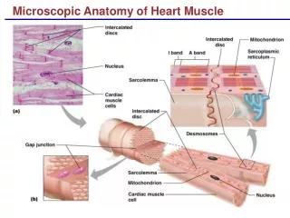
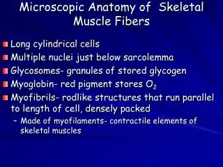
Microscopic AnatomyModifications for Absorption • Small intestine is highly adapted for nutrient absorption • Length provides a huge surface area and its wall has three structural modifications that amplify its absorptive surface enormously: • Plicae circulares • Villi • Microvilli • Most absorption occurs in the proximal part of the small intestine, so these specializations decrease in number toward its distal end
Microscopic AnatomyPlicae Circulares • Circular folds that are deep, permanent folds of the mucosa and submucosa (a) • These folds force chyme to spiral through the lumen, slowing its movement and allowing time for full nutrient absorption
Microscopic AnatomyVilli • Fingerlike projections of the mucosa (a) (b) • Give the mucosa a velvety texture • Epithelial cells of the villi are chiefly absorptive columnar cells • In the core of each villus is a dense capillary bed and a wide lymph capillary called a lacteal
Microscopic AnatomyVilli • Digested food is absorbed through the epithelial cells into both the capillary blood and the lacteal • A slit of smooth muscle (b) (muscularis mucosae) in the villus core allows it to alternately shorten and lengthen, pulsations that: • 1. Increase the contact between the villus and the contents of the intestinal lumen, making absorption more efficient • 2. Milk lymph along through the lacteals
Microscopic AnatomyMicrovilli • Tiny projections of the plasma membrane of the absorptive cells of the mucosa (b) (c) • Gives the mucosal surface a fuzzy appearance called the brush border • The plasma membrane of the microvilli bear enzymes referred to as brush border enzymes, which complete the digestion of carbohydrates and proteins in the small intestine
Histology of the Wall • The four tunics typical of the GI tract (mucosa, submucosa, muscularis externa, adventita) are also seen here, but the mucosa and submucosa are modified to reflect the intestine’s functions in the digestive pathway
Histology of the Wall • Epithelium of the mucosa is largely simple columnar absorptive cells bound by tight junctions and richly endowed with microvilli • Many secretory cells: • Goblet cells: mucus-secreting (d) • Enterogastrones: secretes • Secretin: • A hormone that stimulates sodium bicarbonate secretion by the pancreas and bile secretion by the liver • Decreases gastrointestinal peristalsis and motility • Cholecystokinin: • A hormone that stimulates contraction of the gallbladder and pancreatic secretions • Intraepithelial lymphocytes (T cells): represent an important immunological compartment • Generated locally (not in the Thymus)
Histology of the Wall • Between the villi, the mucosa is studded with pits that lead into tubular intestinal glands called intestinal crypts (crypts of Lieberkuhn): • The epithelial cells that line these crypts secrete intestinal juice: • A watery mixture containing mucus that serves as a carrier fluid for absorbing nutrients from chyme • Deep in the crypts are specialized secretory cells called Paneth cells: • Release lysozyme, an antibacterial enzyme
Histology of the Wall • Submucosa is typical areolar connective tissue • Contains both individual and aggregated lymphoid follicles (Peyer’s patches) • In the duodenal (Brunner’s) glands: (b) • Produce an alkaline (bicarbonate-rich) mucus that helps neutralize the acidic chyme moving in from the stomach • Muscularis is typical and bilayered • Adventitia: covered by visceral peritoneum (serosa)
Intestinal Juice: Composition and Control • Major stimulus is distension or irritation of the intestinal mucosa by hypertonic or acidic chyme • Slightly alkaline (7.4-7.8) • Isotonic with blood plasma • Largely water but it also contains some mucus, which is secreted both by the duodenal glands and by goblet cells of the mucosa • Relatively enzyme-poor because intestinal enzymes are largely limited to the bound enzymes of the brush border
Associated StructuresLiver and Gallbladder • Liver: one of the body’s most important organs, has many metabolic and regulatory roles • It’s digestive function is to produce bile for export to the duodenum: • Bile is a fat emulsifier; that is, it breaks up fats into tiny particles so that they are more accessible to digestive enzymes • Also processes nutrient-laden venous blood delivered to it from the digestive organs, this is a metabolic rather than a digestive role • Gallbladder is chiefly a storage organ for bile
LIVER • Largest gland in the body • Occupies most of the right hypochondriac and epigastric regions • Located under the diaphragm • Lies almost entirely within the rib cage, which provides some protection
LIVER • Has four primary lobes • A mesentery, separates the right and left lobes anteriorly and suspends the liver from the diaphragm and anterior abdominal wall • Entire liver is enclosed by the visceral peritoneum
LIVER • Bile leaves the liver through several bile ducts that ultimately fuse to form the large common hepatic duct, which travels downward toward the duodenum • Along its course, it fuses with the cystic duct draining the gallbladder to form the bile duct
LIVER • The liver is composed of liver lobules, each lobule is a roughly hexagonal (six-sided) structure consisting of plates of liver cells (hepatocytes), organized like bricks in a garden wall • The hepatocyte plates radiate outward from a central vein running in the longitudinal axis of the lobule
LIVER • The liver’s main function is to filter and process the nutrient-rich blood delivered to it • At the end of the six corners of a lobule is a portal triad (three structures) • Branch of the hepatic artery: supplying oxygen-rich arterial blood to the liver • Branch of the hepatic portal vein: carrying venous blood laden with nutrients from the digestive viscera • Bile duct
LIVER • Between the hepatocyte plates are enlarged, leaky capillaries, the liver sinusoids • Blood from both the hepatic portal vein and the hepatic artery percolates from the triad regions through these sinusoids and empties into the central vein • From the central veins blood eventually enters the hepatic veins, which drain the liver, and empty into the inferior vena cava
LIVER • Forming part of the sinusoid walls are star-shaped hepatic macrophages (Kupffer cells)(d) • Remove debris such as bacteria and worn-out blood cells from the blood as it flows past
LIVER • The versatile hepatocytes have large amounts of both rough and smooth ER, Golgi apparatuses, peroxisomes, and mitochondria • Thus equipped, the hepatocytes not only produce bile but can also: • 1.Process the bloodborne nutrients in various ways • Store glucose as glycogen • Use amino acids to make plasma proteins • 2.Store fat-soluble vitamins • 3.Play important roles in detoxification, such as ridding the blood of ammonia by converting it to urea • Blood leaving the liver contains fewer nutrients and waste materials than the blood that entered it
HOMEOSTATIC IMBALANCE • Hepatitis: inflammation of the liver (most often due to viral infection • Hepatitis A virus (HVA): • Transmitted enterically (pertaining to the small intestine) • Sewage contaminated food, raw shellfish, water, feces-mouth route • Infection is self-limiting • 32% of hepatitis cases • Benign form • Frequently in day care centers • Hepatitis B virus (HVB): • Transmitted via blood, contaminated needles, sexual contact • Linked to chronic hepatitis and liver cirrhosis • 40% of hepatitis cases • Elevated risk of cancer • vaccine • Hepatitis C virus (HVC): • Transmitted via blood • Linked to chronic hepatitis and liver cirrhosis • Most important liver disease in the U.S. because it produces persistent or chronic liver infections
HOMEOSTATIC IMBALANCE • Hepatitis D virus (HVD): • Mutated virus that needs HVB to be infectious • Hepatitis E virus (HVE): • Transmitted enterically (pertaining to the small intestine) • Sewage contaminated food, raw shellfish, water, feces-mouth route • Infection is self-limiting • Waterborne epidemics in developing countries • Major case of death in pregnant women • Hepatitis F virus (HVF): • Little is known
HOMEOSTATIC IMBALANCE • Nonviral causes of acute hepatitis include drug toxicity and wild mushroom poisoning • Cirrhosis: • Diffuse and progressive chronic inflammation of the liver that typically results from chronic alcoholism or severe chronic hepatitis • Alcohol-poisoned or damaged hepatocytes regenerate, but the liver’s connective (scar) tissue regenerates faster • As a result, the liver becomes fatty and fibrous and its activity is depressed • As the scar tissue shrinks, it obstructs blood flow throughout the hepatic portal system, causing portal hypertension
COMPOSITION of BILE • Bile is a yellow-green, alkaline solution containing bile salts, bile pigments (primarily bilirubin), cholesterol, neutral fats, phospholipids (lecithin and others), and a variety of electrolytes • Of these only bile salts and phospholipids aid the digestive process • Bile salts: • Primarily cholic acid and chenodeoxycholic acids • Cholesterol derivatives • Role is to emulsify fats—that is, to distribute them throughout the watery intestinal contents • As a result, large fat gobules entering the small intestine are physically separated into millions of small fatty droplets that provide large surface areas for the fat-digesting enzymes to work on • Facilitate fat and cholesterol absorption and help solubilize cholesterol • Although many substances secreted in bile leave the body in feces, bile salts are not among them • Instead, bile salts are conserved by means of a recycling mechanism, called the enterohepatic circulation, and returned to the liver by means of the hepatic portal blood after being reabsorbed by the ileum
COMPOSITION of BILE • Chief bile pigment is bilirubin, a waste product of the heme of hemoglobin formed during the breakdown of worn-out erythrocytes • Absorbed by the liver cells, excreted into bile, and metabolized in the small intestine by resident bacteria • One of the breakdown products, urobilinogen, gives feces a brown color • In the absence of bile, feces are gray-white in color and have fatty streaks (because essentially no fats are digested or absorbed) • Stimulus of bile secretion: • Bile salts • Secretin (released by the intestinal cells)
Gallbladder • Thin-walled green muscular sac • Size of a kiwi fruit • Stores and concentrates bile (absorbing some of its water and ions) that is not needed immediately for digestion • When its muscular wall contracts, bile is expelled into its duct, the cystic duct, and then flows into the bile duct
Regulation of Bile Release into the Small Intestine • When no digestion is occurring, the hepatopancreatic sphincter (guarding the entry of bile and pancreatic juice into the duodenum) is closed and the released bile backs up the cystic duct into the gallbladder, where it is stored until needed • Although the liver makes bile continuously , bile does not usually enter the small intestine until the gallbladder contracts • Major stimulus for gallbladder contraction is when stimulated by cholecystokinin (CCK), an intestinal hormone • Released into the blood when acidic, fatty chyme enters the duodenum • Stimulates the secretion of pancreatic juice • Relaxes the hepatopancreatic sphincter so that bile and pancreatic juice can enter the duodenum • Parasympathetic impulses delivered by the vagus nerves are a minor stimulus for gallbladder contraction
HOMEOSTATIC IMBALANCE • Bile is the major vehicle for cholesterol excretion from the body, and bile salts keep the cholesterol dissolved within bile • Too much cholesterol or too few bile salts leads to cholesterol crystallization, forming gallstones (biliary calculi) which obstruct the flow of bile from the gallbladder • When the gallbladder or its duct contracts, the sharp crystals cause agonizing pain that radiates to the right thoracic region • Treatment: • Ultrasound: pulverization (lithotripsy) • Lasers: vaporization • Surgically removal
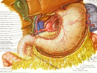
PANCREAS • Is retroperitoneal and lies deep to the greater curvature of the stomach • Pancreatic juice (exocrine product) consists mainly of water and contains enzymes (delivered to the duodenum) that break down all categories of foodstuffs and electrolytes • Pancreatic duct generally fuses with the bile duct just as it enters the duodenum • A smaller accessory pancreatic duct empties directly into the duodenum just proximal to the main duct
PANCREAS • Acini: cluster of secretory cells surrounding ducts • Full of rough endoplasmic reticulum and exhibit deeply staining zymogen granules containing the digestive enzymes they manufacture • Scattered amidst the acini are the more lightly staining pancreatic islets (islets of Langerhans) • Release hormones: insulin and glucagon
Pancreatic Juice • Consists mainly of water, and contains enzymes and electrolytes (primarily bicarbonate ions—making it alkaline-pH 8) • Acinar cells produce the enzyme-rich component of pancreatic juice • The high pH (8) enables it to neutralize acid chyme entering the duodenum and provides the optimal environment for activity of intestinal and pancreatic enzymes
Pancreatic Juice • Pancreatic proteases (protein-digesting enzymes) are produced and released in inactive forms, which are activated in the duodenum, where they do their work (This prevents the pancreas from self-digestion) • Trypsinogen is activated to trypsin by enterokinase (an intestinal brush border enzyme) • Proteases (procarboxypeptidase and chymotrypsinogen) are activated to their active forms (carboxypeptidase and chymotrypsin) by trypsin • Amylase, lipases, and nucleases—are secreted in active form, but require that ions or bile be present in the intestinal lumen for optimal activity
Regulation of Pancreatic Secretion • Regulated by: • Intestinal hormones: • Both act on the pancreas • Secretin released in response to the presence of HCl in the intestine targets the duct cells, prompting their release of watery bicarbonate-rich pancreatic juice • Cholecystokinin (CCK) released in response to the entry of proteins and fats, stimulates the acini to release enzyme-rich pancreatic juice • Parasympathetic nervous system: • Vagal stimulation causes release of pancreatic juice primarily during the cephalic and gastric phases of gastric secretion
Regulation of pancreatic juice secretion by hormonal and neural factors
Digestive Processes Occurring in the Small Intestine • Food reaching the small intestine is far from being digested chemically • Carbohydrates and proteins are partially degraded, but virtually no fat digestion has occurred to this point • Food takes 3 to 6 hours to complete its digestive path through the small intestine, the site of virtually all nutrient absorption
Requirements for Optimal Intestinal Digestive Activity • Although the primary functions of the small intestine are digestion and absorption, intestinal juice provides little of what is needed to perform these functions: • Most substances required for chemical digestion within the small intestine are imported from the pancreas and the liver • Hence, anything that impairs liver or pancreatic function or delivery of their juices to the small intestine severely hinders our ability to digest food and absorb nutrients • Optimal digestive activity in the small intestine depends on a slow, measured delivery of chyme from the stomach (controlled by the pumping action of the stomach pylorus): • Why is this so? • Entering chyme is usually hypertonic • Thus, if large amounts of chyme were rushed into the small intestine, the osmotic water loss from the blood into the intestinal lumen would result in dangerously low blood volume • Additionally, the low pH of entering chyme must be adjusted upward and the chyme must be well mixed with bile and pancreatic juice for digestion to continue
Motility of the Small Intestine • Intestinal smooth muscle mixes chyme thoroughly with bile and pancreatic and intestinal juices, and moves food residues through the ileocecal valve into the large intestine (colon) • In contrast to the peristaltic waves of the stomach, which both mix and propel food, segmentation is the most common motion of the small intestine • Chyme is moved backward and forward in the lumen a few centimeters at a time by alternating contraction and relaxation of rings of smooth muscle • Like the peristalsis of the stomach, segmentation is initiated by intrinsic pacemaker cells in the longitudinal smooth muscle layer (b)
Motility of the Small Intestine • Intensity of segmentation is altered by long and short reflexes (which parasympathetic activity enhances and sympathetic activity decreases) and hormones • True peristalsis occurs only after most nutrients have been absorbed: • At this point, segmenting movements wane, and peristaltic waves initiated in the duodenum begin to sweep slowly along the intestine • Each successive wave is initiated a bit more distally, and this pattern of peristaltic activity is called the migrating mobility complex
Motility of the Small Intestine • Most of the time, the ileocecal sphincter is constricted and closed • However, two mechanisms—one neural and the other hormonal—cause it to relax and allow food residues to enter the cecum when ileal mobility increases • Neural: enhanced activity of the stomach initiates the gastroileal reflex that enhances the force of segmentation • Hormonal: gastrin released by the stomach increases the motility of the ileum and relaxes the ileocecal sphincter • Once the chyme has passed through, it exerts backward pressure that closes the valve’s flaps, preventing regurgitation into the ileum