Download
1 / 11
110 likes | 300 Views
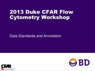
Einstein Flow Cytometry Core Facility 2013 CFAR Retreat, January 15, 2013. Steven A. Porcelli, MD Scientific Director 416 Forchheimer, x3228 steven.porcelli@einstein.yu.edu. FACS Core Laboratory 2004 - 2013 Main location: 3 rd Floor Chanin Building.

E N D
Einstein Flow Cytometry Core Facility 2013 CFAR Retreat, January 15, 2013 Steven A. Porcelli, MD Scientific Director 416 Forchheimer, x3228 steven.porcelli@einstein.yu.edu
FACS Core Laboratory 2004 - 2013 Main location: 3rd Floor Chanin Building Recent renovations funded by $850,000 ARRA Award Supplement to AECC. Completed May 2012 Also: PRICE CENTER (1st Floor) – FACS Core Satellite Laboratory (incorporated 2009)
FACS Core Staff - 2013 Jinghang Zhang, MD Operations Director, 2009- Asst. Operations Director, 2006-2009 Lydia Tesfa, PhD Asst. Operations Director, 2009- Olisambu Uche, PhD Asst. Operations Director, 2011- Grace Wang, B.S. Facility Technician, 2012-
Analyzers 2013 Partec CyCube 6 (Chanin 309) FACSCanto II (Price 159A) LSR II (Chanin 308A and 307A) FACSCalibur DxP10 (Chanin 309)
Analyzers 2013 COMPLEXITY / COST
Analyzers – 2013 The Compucyte iCys Laser Scanning Cytometer (LSC) Similar to a flow cytometer except without the flow component. Cells are analyzed while attached to slides or culture vessels by moving them through the laser with an automated nanostep microscope stage. Although slower than flow cytometry, this device gathers both microscope images and quantitative fluorescence signals on each cell.
Quantification of phagocytosis and cell cycle phase in macrophages by LSC Coelho C & Casadevall A et al. Infect. Immun. 2012;80:1467-1478
Cell Sorters - 2013 Two instrument platforms: MoFlo (Beckman-Coulter) Fastest (triggering rate >70,000 events/second) Most flexible Best for delicate cells and sorting rare events FACSAria II (BD Biosciences) Fast, but not as fast as MoFlo Good configuration, but less flexible Simpler operation Best for applications involving biocontainment
APPROACH TO DELIVERY OF SERVICES Strong emphasis on training facility users to do their own work using Facility resources. For most analytical FACS procedures: We train you to use the instruments and provide guidance as needed. There are exceptions from non-laboratory based researchers; e.g, CD4 counts by MultiSet, priced very low ($29/sample) For cell sorting: We usually do it for you (i.e., you prepare the samples and bring them to us to be sorted according to your specifications) There is an option for self operation of FACSAria for non-biohazard related sorting
Costs for services – Charge back rates (mostly unchanged since 2007) 20% off these rates for CFAR 20% discounts on all services also applied to CFAR and Diabetes Center members