Download
1 / 38
380 likes | 522 Views
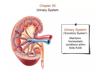
Chapter 28 Urinary System. Overview of the Urinary System. Kidneys— principal organs of the urinary system accessory organs are ureters, urinary bladder, and urethra

E N D
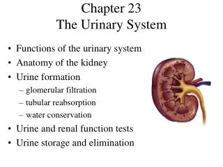
Chapter 28Urinary System Slide
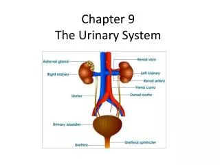
Overview of the Urinary System • Kidneys— principal organs of the urinary system • accessory organs are ureters, urinary bladder, and urethra • Urinary system— regulates the content of blood plasma to maintain homeostasis of the internal fluid environment within normal limits • “blood plasma balancer” Slide
Mink Slide
Human Slide
Gross structure • Kidneys (two) • Roughly oval with a medial indentation • Left kidney often larger than right • Right kidney is a little lower • Lie on either side of the vertebral column between T12 and L3 • Heavy cushion of fat surrounds each kidney Slide
Internal structures of kidney • Cortex and medulla • Renal pyramids comprise much of the medullary tissue • Renal columns— where cortical tissue dips into the medulla between the pyramids • Calyx (KAY liks)— cuplike structure at each renal papilla to collect urine • join together to form renal pelvis • Renal pelvis narrows as it exits kidney to become ureter Slide
Gross structure • Renal artery— large branch of abdominal aorta; brings blood into each kidney • Kidneys are HIGHLY vascular • Every minute 1,200 mL blood flows through them • 1/5 of all blood pumped by heart goes to the kidneys Slide
Gross structure FYI: Pattern of blood flow through kidneys—abdominal aorta → renal artery → segmental arteries → lobar arteries → interlobar arteries → arcuate arteries → interlobular artery → afferent arteriole → glomerulus (glomerular capillaries) → efferent arteriole → peritubular capillaries (vasa recta) → interlobular veins → arcuate veins → interlobar veins → lobar veins → segmental veins → renal vein → inferior vena cava (Figure 28-3, Page 831) Slide
Gross structure Juxtaglomerular apparatus (juks-tah-glo-MER-yoo-lar) • located near the glomerulus • Helps regulate blood pressure by secreting renin when blood pressure in afferent arteriole drops Ureter • tube running from each kidney to urinary bladder • composed of three layers • mucous lining • muscular middle layer • fibrous outer layer Slide
Gross structure • Urinary bladder • collapsible bag located behind the symphysis pubis made mostly of smooth muscle tissue • can distend considerably • Functions • Reservoir for urine before it leaves the body • Aided by the urethra, expels urine from the body • Mechanism for voiding • Voluntary relaxation of external sphincter muscle • Regions of the detrusor muscle contract reflexively • Urine is forced out of the bladder and through the urethra Slide
Gross structure Urethra • Small mucous membrane–lined tube • In females • lies posterior to symphysis pubis and anterior to vagina • approximately 3 cm long • In males • after leaving the bladder, passes through prostate gland where it is joined by two ejaculatory ducts • from prostate, extends to base of penis and then through center of penis, and ends as urinary meatus • approximately 20 cm long • male urethra is part of the urinary system, as well as part of the reproductive system Slide
Nephrons • Nephrons, the microscopic functional units, make up the bulk of the kidney • those located in renal cortex called cortical nephrons • those near junction of cortical and medullary layers called juxtamedullary nephrons Slide
Structure of the Nephron Renal corpuscle • Bowman’s capsule— cup-shaped mouth of nephron • Glomerulus— network of fine capillaries in Bowman’s capsule • together called renal corpuscle • located in cortex of kidney • Glomerular-capsular membrane • function is filtration Slide
Structure of the Nephron • Proximal tubule • first part of renal tubule nearest to Bowman’s capsule • also known as proximal convoluted tubule Loop of Henle • Consists of a thin descending limb, a sharp turning, and a thick ascending limb Cortical nephron • a nephron with a loop of Henle that does not dip into the medulla but remains almost entirely within the cortex • constitute about 85% of total nephron numbers Slide
Structure of the Nephron Distal tubule • convoluted tubule beyond the loop of Henle • also known as distal convoluted tubule Collecting duct • Straight tubule joined by the distal tubules of several nephrons • Joins larger ducts • larger collecting ducts of one renal pyramid converge to form one tube that opens at a renal papilla into a calyx Slide
Kidney Function • Chief functions of kidney are to process blood and form urine • Basic functional unit of kidney is nephron • Forms urine through three processes • Filtration— movement of water and protein-free solutes from plasma in glomerulus into space of Bowman’s capsule • Tubular reabsorption— movement of molecules out of tubule and into peritubular blood • Tubular secretion— movement of molecules out of peritubular blood and into tubule for excretion Slide
1: Filtration • Filtration— 1st step in blood processing that occurs in the renal corpuscles • From blood in the glomerular capillaries, about 180 liters of water and solutes filter into Bowman’s capsule each day • takes place through the glomerular-capsular membrane • Filtration occurs as a result of existence of a pressure gradient • Glomerular capillary filtration occurs rapidly as a result of the increased number of fenestrations (openings) • Glomerular pressure and filtration are directly related to systemic blood pressure Slide
2: Reabsorption • Reabsorption- 2nd step • occurs as a result of passive and active transport mechanisms from all parts of the renal tubules • major portion of reabsorption occurs in proximal tubules • Reabsorption in proximal tubule: most water and solutes are recovered by the blood, leaving only a small volume of tubule fluid to move on to the loop of Henle Slide
2: Reabsorption Reabsorption in proximal tubule • Sodium— actively transported out of tubule fluid and into blood • Glucose and amino acids— passively transported out of tubule fluid by means of the sodium cotransport mechanism • Chloride, phosphate, and bicarbonate ions passively move into blood because of an imbalance of electrical charge • Water— movement of sodium and chloride into blood causes an osmotic imbalance, moving water passively into blood • Urea— approximately one half of urea passively moves out of tubule with the remaining urea moving on to the loop of Henle Slide
2: Reabsorption Reabsorption in the loop of Henle • Water is reabsorbed from the tubule fluid • urea is picked up from the interstitial fluid in the descending limb • Sodium and chloride are reabsorbed from the filtrate in the ascending limb, where the reabsorption of salt makes the tubule fluid dilute and creates and maintains a high osmotic pressure of the medulla’s interstitial fluid • Go to this Great Website! Slide
2: Reabsorption • Reabsorption in the distal tubules and collecting ducts • Distal tubule reabsorbs sodium by active transport but in smaller amounts than in the proximal tubule • ADH is secreted by the posterior pituitary and targets the cells of distal tubules and collecting ducts to make them more permeable to water • With the reabsorption of water in the collecting duct, [urea] of tubule fluid increases urea to diffuse out of collecting duct into medullary interstitial fluid • Urea participates in a countercurrent multiplier mechanism that, along with countercurrent mechanisms of the loop of Henle and vasa recta, maintains the high osmotic pressure needed to form concentrated urine and avoid dehydration Slide
3: Secretion • H +, K+ and NH3 are actively transported out of the blood into filtrate to be excreted. This increase in concentration increases the activity of the Na+/K+ pump in the distal and collecting tubule. Slide
3: Secretion • Tubular secretion— the movement of substances out of blood and into tubular fluid • Descending limb of loop of Henle secretes urea via diffusion • Distal and collecting tubules secrete potassium, hydrogen, and ammonium ions • Aldosterone— hormone that targets the cells of distal and collecting tubule cells, causes increased activity of sodium-potassium pumps increasing [Na+] • Secretion of hydrogen ions increases with decreases blood [H+] Slide
Regulation of urine volume • Antidiuretic hormone (ADH) influences water reabsorption • as water is reabsorbed, total volume of urine is reduced by amount of water removed by tubules • ADH reduces water loss • Aldosterone • secreted by adrenal cortex • increases distal tubule absorption of sodium, raising [Na+] of blood • thus promoting reabsorption of water • Atrial natriuretic hormone (ANH) • secreted by specialized atrial muscle fibers (in atria) • promotes loss of sodium via urine • opposes aldosterone, causing the kidneys to reabsorb less water • thereby produce more urine • This reduces blood pressure by reducing the amount of water in blood Slide
Regulation of urine volume • Tubuloglomerular feedback mechanism maintains constant glomerular filtration rate (GFR) by regulating resistance in afferent arterioles • protects kidney GFR function from rapid blood pressure variations • dependent on juxtaglomerular apparatus • Myogenic mechanism— rapid and effective regulation of GFR via changes in afferent arteriole smooth muscle contraction and relaxation • Urine volume— also related to total amount of solutes other than sodium excreted in the urine • generally, the more solutes, the more urine Slide
Urine Composition • approximately 95% water with several substances dissolved in it • the most important are the following: • Nitrogenous wastes— result of protein metabolism • urea, uric acid, ammonia, and creatinine • Electrolytes • sodium, potassium, ammonium, chloride, bicarbonate, phosphate, and sulfate • amounts and kinds of minerals vary with diet and other factors • Toxins— during disease, bacterial poisons leave the body in urine • Pigments— especially urochromes (make it yellow) • Hormones— high hormone levels may spill into the filtrate • Abnormal constituents— such as blood, glucose, albumin, protein, WBC Slide
Urine dip stick Slide
The Big Picture: Urinary System and the Whole Body • Homeostasis of water and electrolytes in body fluids relies on proper functioning of the kidneys • Nephrons process blood to adjust its content to maintain a relatively constant internal environment • Urinary and cardiovascular systems are interdependent • Endocrine and nervous systems must operate properly to ensure efficient kidney function Slide