Download
1 / 6
60 likes | 72 Views
Tuberculosis IgG ELISA Kit can be provided from Creative Diagnostics.t<br>https://www.creative-diagnostics.com/Tuberculosis-IgG-EIA-Kit-107207-466.htm<br>

E N D
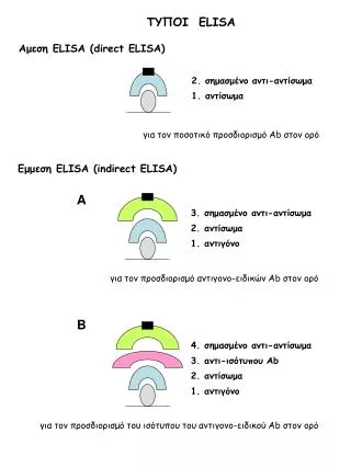
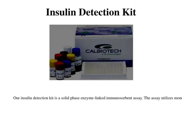
Tuberculosis IgG ELISA Kit Cat. No.:DEIA1023 Pkg.Size:96T Intended use This kit is an enzyme-linked immunosorbent assay for qualitative detection of IgG antibodies to Mycobacterium tuberculosis in human serum or plasma. It is intended for diagnosing and monitoring of patients related to infection by M. tuberculosis and other Mycobacteria. General Description It has been estimated that more than 1.7 million people were infected with Mycobacterium tuberculosis (MTb) in 1990 and this number increase every year due to new infections (HIV) and immigration. Now, with worldwide prevalence of 30 million cases and an incidence of 10 million new cases each year, tuberculosis (TB) is one of the most important health threatening problems worldwide. IgG antibodies to MTb are routinely investigated in chronic infections and allow monitoring and management of the patients during treatment because inefficient therapy normally is characterized by increasing of the synthesis and excretion IgG. On the other hand, low levels of IgG in an inactive disease state indicates the non-evacuation of intra cellular infectious pockets with possible progression of the disease to active at later state. Principle Of The Test This kit employs solid phase, indirect ELISA assay for detection of IgG antibodies to MTb in two-step incubation procedure. Polystyrene microwell strips are pre-coated with affinity purified MTb antigens including A60 antigen complex. During the first incubation step, MTb IgG specific antibodies, if present, will be bound to the solid phase pre-coated antigen complexes. The wells are washed to remove unbound serum proteins, and rabbit anti-human IgG antibodies (anti-IgG) conjugated to horseradish peroxidase (HRP) are added. During the second incubation step, these HRP-conjugated antibodies will be bound to any antigen- IgG complexes previously formed and the unbound HRP-conjugate is then removed by washing. Chromogen solutions containing Tetramethylbenzidine (TMB) and urea peroxide are added to the wells. In presence of the antigen-(IgG)-anti-IgG (HRP) immunocomplex, the colorless Chromogens are hydrolyzed by the bound HRP conjugate to a blue colored product. The blue color turns yellow after stopping the reaction with sulfuric acid. The amount of color intensity can be measured and is proportional to the amount of antibody captured in the wells, and to the sample respectively. Wells containing samples negative for IgG antibodies to MTb remain colorless. Reagents And Materials Provided MICROWELL PLATE: 1 plate Blank microwell strips fixed on white strip holder. The plate is sealed in aluminum pouch with desiccant. 8×12/12×8-well strips per plate. Each well contains affinity purified MTb antigens. The microwell strips can be broken to be used separately. Place unused wells in the plastic sealable storage bag together with the desiccant and return at 2~8℃. NEGATIVE CONTROL: 1 vial Colorless liquid filled in vial with green screw cap 0.5ml per vial. Protein-stabilized buffer tested non-reactive for MTb IgG antibodies. Preservatives: 0.1% Proclin 300. Ready to use as supplied. Once open, stable for one month at 2-8℃. POSITIVE CONTROL SERUM: 1 vial Creative Diagnostics. All rights reserved 45-16 Ramsey Road Shirley, NY 11967, USA Tel: 631-624-4882 ·Fax:631-614-7828 E-mail: info@creative-diagnostics.com www.creative-diagnostics.com
Red liquid filled in vial with red screw cap 0.5ml per vial. MTb antibodies diluted in protein-stabilized buffer containing preservatives: 0.1% Proclin 300. Ready to use as supplied. Once open, stable for one month at 2-8℃. SPECIMEN DILUENT: 1 vial Blue liquid filled in a white vial with blue screw cap. 12ml per vial Protein-stabilized buffer, casein, and sucrose solution. Ready to use as supplied. Once open, stable for one month at 2-8℃. HRP-CONJUGATE REAGENT: 1 vial Red liquid filled in a white vial with red screw cap. 12ml per vial. Horseradish peroxidase-conjugated rabbit anti-human IgG antibodies. Ready to use as supplied. Once open, stable for one month at 2-8℃. STOCK WASH BUFFER: 1 bottle DILUTE BEFORE USE Colorless liquid. 50 ml per bottle. PH 7.4, 20 × PBS (Containing Tween 20 as a detergent) The concentrate must be diluted 1:20 with distilled/deionized water before use. Once diluted, stable for one weeks at room temperature or for two weeks at 2-8℃. CHROMOGEN SOLUTION A: 1 vial Colorless liquid filled in white vial with green screw cap. 6 ml per vial. Urea peroxide solution. Ready to use as supplied. Once open, stable for one month at 2-8℃. CHROMOGEN SOLUTION B: 1 vial Colorless liquid filled in the black vial with black screw cap. 6 ml per vial. TMB solution- Tetramethylbenzidine dissolved in citric acid. Ready to use as supplied. Once open, stable for one month at 2-8℃. STOP SOLUTION: 1 vial Colorless liquid filled in white vial with yellow screw cap. 6 ml per vial. Diluted sulfuric acid solution (2.0M H2SO4). Ready to use as supplied. PLASTIC SEALABLE BAG: 1 unit For enclosing the strips not in use. CARDBOARD PLATE COVER: 2 sheets To cover the plates during incubation and prevent evaporation or contamination of the wells. PACKAGE INSERTS: 1 copy Creative Diagnostics. All rights reserved 45-16 Ramsey Road Shirley, NY 11967, USA Tel: 631-624-4882 ·Fax:631-614-7828 E-mail: info@creative-diagnostics.com www.creative-diagnostics.com
Materials Required But Not Supplied 1. Freshly distilled or deionized water. 2. Disposable gloves and timer. 3. Appropriate waste containers for potentially contaminated materials. 4. Disposable V-shaped troughs. 5. Dispensing system and/or pipette (single or multichannel), disposable pipette tips. 6. Absorbent tissue or clean towel. 7. Dry incubator or water bath, 37±0.5℃. 8. Microshaker for dissolving and mixing conjugate with samples. 9. Microwell plate reader, single wavelength 450nm or dual wavelength 450nm and 630nm. 10. Microwell aspiration/wash system. Storage The components of the kit will remain stable through the expiration date indicated on the label and package when stored between 2-8 ℃, do not freeze. To assure maximum performance of this TB IgG ELISA kit, protect the reagents from contamination with microorganism or chemicals during storage. Specimen Collection And Handling 1. Sample Collection: Either fresh serum or plasma samples can be used for this assay. Blood collected by venipuncture should be allowed to clot naturally and completely – the serum/plasma must be separated from the clot as early as possible as to avoid hemolysis of the RBC. Care should be taken to ensure that the serum samples are clear and not contaminated by microorganisms. Any visible particulate matters in the sample should be removed by centrifugation at 3000 RPM for at least 20 minutes at room temperature, or by filtration on 0.22u filters. Plasma samples collected into EDTA, sodium citrate or heparin may be tested, but highly lipaemic, icteric, or hemolized samples should not be used as they could give erroneous results in the assay. Do not heat inactivate samples. This can cause sample deterioration. 2. Transportation and Storage: Store samples at 2-8 ℃. Samples not required for assaying within 3 days should be stored frozen (-20℃ or lower).Multiple freeze-thaw cycles should be avoided. For shipment, samples should be packaged and labeled in accordance with the existing local and international regulations for transport of clinical samples and ethological agents. Assay Steps Step1 Reagents preparation: Allow the reagents and samples to reach room temperature (18-30°C) for at least 15-30minutes. Check the Wash buffer concentrate for the presence of salt crystals. If crystals have formed in the solution, resolubilize by warming at 37 ℃ until crystals dissolve. Dilute the stock wash Buffer 1 to 20 with distilled or deionized water. Use only clean vessels to dilute the Wash buffer. Step2 Numbering Wells: Set the strips needed in strip-holder and number sufficient number of wells including three Negative control (e.g. B1, C1, D1), two Positive control (e.g. E1, F1) and one Blank (A1, Neither samples nor HRP-Conjugate should be added into the Blank well). If the results will be determined by using dual wavelength plate reader, the requirement for use of Blank well could be omitted. Use only number of strips required for the test. Step3 Adding Diluent: Add 100μl Specimen Diluent into each well except in the blank. Step4 Adding Sample: Add 10μl of Specimen and 10μl of Positive and Negative controls into their respective wells. Note: Use a separate disposal pipette tip for each specimen, Negative Control and Positive Control as to avoid cross-contamination. Mix by tapping the plate gently. Step5 Incubating: Cover the plate with the plate cover and incubate for 30 minutes at 37°C. It is recommended to use water tank to assure the temperature stability and humidity during incubation. If dry incubator is used, do not open the door frequently. Step6 Washing: After the end of the incubation, remove and discard the plate cover. Wash each well 5 times with diluted Wash Creative Diagnostics. All rights reserved 45-16 Ramsey Road Shirley, NY 11967, USA Tel: 631-624-4882 ·Fax:631-614-7828 E-mail: info@creative-diagnostics.com www.creative-diagnostics.com
buffer. Each time allow the microwells to soak for 30-60 seconds. After the final washing cycle, turn the strips plate onto blotting paper or clean towel, and tap the plate to remove any remainders. Step7 Adding HRP-Conjugate: Add 100 μl HRP-Conjugate to each well except the Blank. Step8 HRP-Conjugate Incubating: Cover the plate with the plate cover and incubate for 30 minutes at 37°C. Step9 Washing: After the end of the incubation, remove and discard the plate cover. Wash each well 5 times with diluted Wash buffer as in Step6. Step10 Coloring: Dispense 50μl of Chromogen A and 50μl Chromogen B solution into each well including the Blank and mix by tapping the plate gently. Incubate the plate at 37℃ for 15 minutes avoiding light. The enzymatic reaction between the Chromogen A/B solutions produces blue color in Positive control and TB IgG Positive sample wells. Step11 Stopping Reaction: Using a multichannel pipette or manually add 50 μl Stop Solution into each well and mix by tapping the plate gently. Intensive yellow color develops in Positive control and TB IgG Positive sample wells. Step12 Measuring the Absorbance: Calibrate the plate reader with the Blank well and read the absorbance at 450nm. If a dual filter instrument is used, set the reference wavelength at 630nm. Calculate the Cut-off value and evaluate the results. (Note: read the absorbance within 5 minutes after stopping the reaction) Quality Control The test results are valid if the Quality Control criteria are verified. It is advisable that each laboratory must establish appropriate quality control system with quality control material similar to or identical with the patient sample being analyzed. 1. The OD value of the Blank well, which contains only Chromogens and Stop solution, is less than 0.080 at 450 nm. 2. The OD value of the Negative control must be equal to or greater than 0.800 at 450/630nm or at 450nm after blanking. 3. The OD value of the Positive control must be less than 0.100 at 450/630nm or at 450nm after blanking. Calculation Each microplate should be considered separately when calculating and interpreting the results, regardless of the number of plates concurrently processed. The results are calculated by relating each sample’s optical density (OD) value to the Cut-off value (C.O.) of the plate. If the Cut-off reading is based on single filter plate reader, the results should be calculated by subtracting the Blank well OD value from the print report values of samples and controls. In case the reading is based on dual filter plate reader, do not subtract the Blank well OD from the print report values of samples and controls. Calculation of Cut-off value (C.O.) = *Nc + 0.15 *Nc = the mean absorbance value for three negative controls. If one of the Negative control values does not meet the Quality Control Range specifications, it should be discarded and the mean value is calculated again using the remaining two values. If more than one control OD value does not meet the Quality control Range specifications, the test is invalid and must be repeated. Interpretation of Results (S = the individual absorbance (OD) of each specimen) Negative Results (S/C.O. <1): samples giving absorbance less than the Cut-off value are negative for this assay, which indicates that no IgG antibodies to Mycobacterium tuberculosis have been detected with this TB IgG ELISA kit. Positive Results (S/C.O.≥1) : samples giving an absorbance greater than or equal to the Cut-off value are considered initially reactive, which indicates that IgG antibodies to Mycobacterium tuberculosis have probably been detected using this TB IgG ELISA kit. Retesting in duplicates of any initially reactive sample is recommended. Repeatedly reactive samples can be considered positive for IgG antibodies to Mycobacteria. Borderline: (S/C.O. =0.9-1.1) Samples with absorbance to Cut-off ratio between 0.9 and 1.1 are considered borderline and retesting of these samples in duplicates is recommended to confirm the results. Repeatedly positive samples can be considered positive for antibodies to MTb. The results from this assay must be used in conjunction with other Mycobacterium tuberculosis serological test results to determine the immunological response to, and the stage of Tuberculosis infection. Creative Diagnostics. All rights reserved 45-16 Ramsey Road Shirley, NY 11967, USA Tel: 631-624-4882 ·Fax:631-614-7828 E-mail: info@creative-diagnostics.com www.creative-diagnostics.com
Specificity Analytical Specificity: 1. No cross reactivity observed with samples from patients infected with HAV, HIV, HBV, HCV, CMV, and TP. 2. No interference was observed from rheumatoid factors up to 2000U/ml. 3. These assay performance characteristics are unaffected from elevated concentrations of bilirubin, hemoglobin, and triolein. 4. Frozen specimens have been tested to check for interferences due to collection and storage. Precautions The ELISA assay is a time and temperature sensitive method. To avoid incorrect result, strictly follow the test procedure steps and do not modify them. 1. Do not exchange reagents from different lots, or use reagents from other commercially available kits. The components of the kit are precisely matched as to achieve optimal performance during testing. 2. Make sure that all reagents are within the validity indicated on the kit box and are of the same lot. Never use reagents beyond the expiry date stated on reagents labels or on the kit box. 3. CAUTION - CRITICAL STEP: Allow the reagents and samples to stabilize at room temperature (18-30 ℃ ) before use. Shake reagent gently before, and return to 2-8 ℃ immediately after use. 4. Use only sufficient volume of sample as indicated in the procedure steps. Failure to do so, may cause in low sensitivity of the assay. 5. Do not touch the bottom exterior of the wells; fingerprints or scratches may interfere with microwell reading. 6. When reading the results, ensure that the plate bottom is dry and there are no air-bubbles inside the wells. 7. Never allow the microplate wells to dry after the washing step. Immediately proceed to the next step. Avoid the formation of air -bubbles when adding the reagents. 8. Avoid assay steps long time interruptions. Assure same working conditions for all wells. 9. Calibrate the pipette frequently to assure the accuracy of samples/reagents dispensing. Always use different disposal pipette tips for each specimen and reagents as to avoid cross-contaminations. Never pipette solutions by mouth. 10. The use of automatic pipettes is recommended. 11. Assure that the incubation temperature is 37℃ inside the incubator. 12. When adding samples, avoid touching the well’s bottom with the pipette tip. 13. When reading the results with a plate reader, it is recommended to determine the absorbance at 450nm or at 450nm with reference at 630nm. 14. All specimens from human origin should be considered as potentially infectious. Creative Diagnostics. All rights reserved 45-16 Ramsey Road Shirley, NY 11967, USA Tel: 631-624-4882 ·Fax:631-614-7828 E-mail: info@creative-diagnostics.com www.creative-diagnostics.com
15. Materials from human origin may have been used in the kit. These materials have been tested with tests kits with accepted performance and found negative for antibodies to HIV ½, HCV, TP and HBsAg. However, there is no analytical method that can assure that infectious agents in the specimens or reagents are completely absent. Therefore, handle reagents and specimens with extreme caution as if capable of transmitting infectious diseases. Strict adherence to GLP (Good Laboratory Practice) regulations can ensure the personal safety. Never eat, drink, smoke, or apply cosmetics in the assay laboratory. 16. Bovine derived sera may have been used in this kit. Bovine serum albumin (BSA) and fetal calf sera (FCS) are derived from animals from BSE/TSE free-geographical areas. 17. The pipette tips, vials, strips and sample containers should be collected and autoclaved for 1hour at 121℃ or treated with 10% sodium hypochlorite for 30minutes to decontaminate before any further steps for disposal. 18. The Stop solution (2M H2SO4 ) is a strong acid. Corrosive. Use it with appropriate care. Wipe up spills immediately or wash with water if come into contact with the skin or eyes. Proclin 300 used as a preservative can cause sensation of the skin. 19. The enzymatic activity of the HRP-conjugate might be affected from dust, reactive chemical, and substances like sodium hypochlorite, acids, alkalins etc. Do not perform the assay in the presence of such substances. 20. Materials Safety Data Sheet (MSDS) available upon request. 21. If using fully automated microplate processing system, during incubation, do not cover the plates with the plate cover. The tapping out of the remainders inside the plate after washing, can also be omitted. Limitations 1. Non-repeatable positive result may occur due to the general biological characteristics of the ELISA method. The assay is designed to achieve very high performance characteristics of sensitivity and specificity and the “indirect” model minimizes the unspecific reactions due to interference with unknown matters in sample and the anti-human IgG antibodies. Antibodies may be undetectable during the early stages of the disease and in some immunosuppressed individuals. 2. Positive results must be confirmed with another available method and must be interpreted together with the patient clinical information and other laboratory results like X-ray and microbiolog 2. Common sources for mistakes are: kits beyond the expiry date, bad washing procedures, contaminated reagents, incorrect assay procedure steps, insufficient aspiration during washing, failure to add samples or reagents, equipment, timing, volumes, sample nature and quality. 3. The prevalence of the marker will affect the assay’s predictive values. 4. If, after retesting of the initially reactive samples, the assay results are negative , these samples should be considered as non- repeatable (false positive) and interpreted as negative. As with many very sensitive ELISA assays, false positive results can occur due to the several reasons, most of which are related but not limited to inadequate washing step. 5. This kit is intended ONLY for testing of individual serum or plasma samples. Do not use it for testing of cadaver samples, saliva, urine or other body fluids, or pooled (mixed) blood. 6. This is a qualitative assay and the results cannot be use to measure antibodies concentrations. REFERENCES 1. Hasløv, K. Boosting effects in BCG-vaccinated guineapigs of tuberculin skin tests on subsequent skin tests and on the in vitro lymphocyte transformation response. 1983. International Symposium on BCG Vaccines and Tuberculins, Budapest, Hungary. Develop. biol. Standard. 58: 517-521 (S. Karger, Basel. 1986). 2. Zou YL,Zhnag JD, Chen MH,Shi GQ,Prignot J,Cocito C (1194) Serological analysis of pulmonary and extrapulmonary tuberculosis with enzyme-linked immunosorbent assay for anti-A60 immunoglobulings C.I.D. 19: 1084-1091 3. Wirrmann C(1990) Public health applications of serological tests for tuberculosis :Study of the incidence of inapparent infectious employees of an alsacian supermarket. Eur J Epidemol 6:304-308 4. Harboe M (1981) Antigens of PPD ,old tuberculin and autoclaved M.bovis BCG studied by cross immunoelectrophoresis .Am Rev Respir Dis 124:80-87 Creative Diagnostics. All rights reserved 45-16 Ramsey Road Shirley, NY 11967, USA Tel: 631-624-4882 ·Fax:631-614-7828 E-mail: info@creative-diagnostics.com www.creative-diagnostics.com