Download
1 / 11
110 likes | 133 Views
Engage students in discussing pathogens, immune response, and diseases through group activities, glossary matching, jigsaw learning, and practical bacterial staining. Enhance understanding of bacteria vs viruses, pathogen identification, and phagocytosis process.

E N D
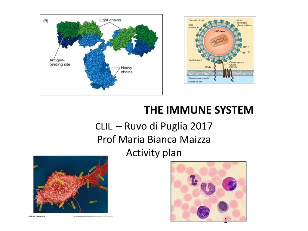
CLIL– Ruvo di Puglia 2017 Prof Maria Bianca Maizza Activityplan THE IMMUNE SYSTEM
ACTIVATING PRIOR KNOWLEDGE Brainstorming: “Do you know....?” I will stimulate a discussion in class: in small groups, the learners have to talk about pathogens and immune response. Then each group discuss with the others, involving the whole class in one unique discussion. ( Pyramid discussion ) Cancer Influenza Measles Heart disease Rubella Mumps AIDS Which of these are infectious diseases? Why do you get ill? What is AIDS? What is the difference between a virus and a bacterium? What is an allergy? What is the purpose of vaccination?
GLOSSARY This step consists in giving a glossary to the students. I will cut out each term and definition separately. I will shuffle the cards. Then the students have to match each term to the correct definition to form a pair of columns on a table. (domino game)
Activity 1TASK-BASED ACTIVITY: BACTERIUM OR VIRUS? Antibiotics are chemicalsubstances, usuallyisolatedfrom fungi, thatinhibitsbacterialgrowth. They are noteffectiveagainstviruses. Beforeadministeringantibioticsto the patients, the doctorshouldknowwhethertheirdiseases are causedby a bacterium or a virus The questionthat I will propose to the studentsis : Can you help the doctor? • The studentswillwatch a video on the bacteria and viruses • www.youtube.com/watch?v=3xRttWuf3wQ
The process of organising a Jigsaw activity Step 1: I divide the classinto‘expert’groupsoffourtosixlearners. Eachgroupchoose a letter or a colour or a name. Theninto the group a numberisassignedtoeachstudent ( 1 to 6 ) Step 2: I giveinformation about 2- 3 pathogenstoeachgroup .In eachgroup the studentsshouldspend some timereading, discussing and helpingeachothertounderstandthe information. Word banks and dictionariesmaybeuseful at this stage. Step 3: Learners, who are becomenow‘experts’ on theirsubjectmoveinto‘jigsaw’groups, forexampleall the number 1 students work in a group, all the number 2 students work in anothergroup and so on. Step 4: Each‘expert’learner in turn shareswiththeir‘jigsaw’group the information theywereoriginallygiven. . Step 5: The ‘jigsaw’ grouptogether complete a task whichrequiresthemtounderstandallof the information sharedbyeach‘expert’. Thiscouldbeanythingthatrequireseachlearnertocontributetheirpieceofknowledge: fillingwords in a grid or a table, completing a diagram, designing a poster....
Jigsaw activity Using textbooks or internet, find useful information on the following pathogens and complete the T chart by writing the name of the bacteria and viruses from the list below • Herpes zoster • Neisseria meningitidis • Streptococcus pneumoniae • Bacillus anthracis • Hepatitis C • Esherichia coli • Streptococcus pyogenes • Herpes simplex • Vibrio cholera • Staphylococcus aureus • HIV bacteria viruses
PRACTICAL ACTIVITY: SHAPE AND STAINING OF BACTERIA • Activity2 in laband • at home : The studentswillsee some slidesaboutbacterialmorphology Example of bacterial morphology
Activity in lab: Gram staining Background: Gram staining is a differential staining based on the physical and chemical properties of bacterial cell wall. Procedure: Using a toothpick, take some bacteria from the solid or liquid culture. Make a smear on a clean microscope slide. Take the slide with the tweezers and briefly pass it on a flame (Bunsen) FIXATION Let the slide cool down at room temperature. Put the slide on a support over a dish. Cover with a few drops of crystal violet 1min PRIMARY STAINING Rinse with water to remove excess staining. Cover with a few drops of lugol 1 min MORDANT Rinse with water. Add some ethanol until you see a grey/blue colour (<30 sec) DECOLORIZATION Rinse with water. Cover with a few drops of safranin 1 min COUNTER STAINING Rinse with water. Let it dry. Observe at the microscope. Record your results (drawings). http://faculty.mc3.edu/jearl/ML/ml-5.htm Result: Gram-positive bacteria are dark-blue stained; Gram-negative bacteria are pink-red stained.
Activity at home: how to distinguish bacteria developing learner’s ability to find information from various sources 1) Makedrawingsto show the chemicalcompositionofGram-positive and Gram-negative cellwall. Gram-positive cell wall Gram-negative cell wall 2) Search information on the internet usefulto complete the followingtable:
Activity 3Listen– define – put in order – perform a practicalactivity – file a report - draw http://highered.mheducation.com/sites/0072495855/student_view0/chapter2/animation__phagocytosis.html • 1) Watch and listento the video: • 2)Define the followingtermsmentioned in the video: • Phagocytes - Surfacereceptors – Opsonization – Phagosome – Lysosome - Exocytosis • 3) Listento the videoagain and put in the correctorder the followingstepsoccurringduring the processofphagocytosis: • The phagocyteattachesto the microorganisms. • Digestedcontentof the phagolysosomeiseliminatedbyexocytosis. • Phagocytes are attractedto the area ofinvasionbychemicalsubstances. • Lysosomes fuse with the phagosomereleasing digestive enzymes. • The microorganismsisthenengulfedby the phagocyteinto a vacuoleknownas a phagosome. • Inside the phagolysosomemicroorganisms are killed and digested.
4)Practicalactivity • Phagocytosisis a very common cellularprocess in nature. • Macrophages and neutrophils in the human body performphagocytosistocapturepathogens. • Some unicellularorganisms, suchasParamecium, usephagocytosistocapturetheir “food”. Watch the video: http://www.youtube.com/watch?v=l9ymaSzcsdY • You can perform the sameexperimentfollowingthis procedure: • place some hay in a beakerfilledwith water. • keepit open forabout a week toget a culture ofciliates, suchasparamecium. • stainyeastcellswith Congo red • mix together the culture ofciliates and the stainedyeast • observe at the microscope • 5) File a written reportdescribingyourobservation. • 6) Makedrawingsof the processofphagocytosisperformedbyParameciumasyouobservedit at the light microscope.


