Download
1 / 53
530 likes | 712 Views
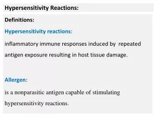
HYPERSENSITIVITY REACTIONS. HYPERSENSITIVITY REACTIONS Innocous materials can cause hypersensitivity in certain individuals. unwanted inflammation damaged cells and tissues. Non-proper reaction of the immune system to foreign substances

E N D
HYPERSENSITIVITY REACTIONS Innocous materials can cause hypersensitivity in certain individuals unwanted inflammation damaged cells and tissues Non-proper reaction of the immune system to foreign substances Mainly harmless substances – after second or multiple times
TYPES OF HYPERSENSITIVITY REACTIONS mostly appear together with autoimmune diseases
ANTIBODY MEDIATED HYPERSENSITIVITY REACTIONS Hay fever Asthma Systemic anaphylaxis Certain drug allergies (penicillin) Serum sickness Arthus reaction
TYPE I HYPERSENSITIVITY REACTION ALLERGY
DIFFERENCES OF IMMUNE RESPONSES INDUCED BY ALLERGENS AND PATHOGENS • PATHOGEN • Deviving • Escape mechanism • DANGER SIGNAL • Microbial DNS, CpG-ODN • dsRNS IFN • LPS (Gram-), PG (Gram+) • HSP • Inflammatory cytokin • TNF-, IL-1 • DC/TISSUE DEMAGE • Activation of innate immunity • Inflammation, neurotransmitters (VIP) • ALLERGEN • Non deviding • Act together with other environmental effects • DANGER SIGNAL • Ni, Mn, Co, Sn chlorides • Ion channel inhibitors • Energy homeostasis • DC/TISSUE DEMAGE • TNCB, DNCB • Der p1 – cisztein proteáz • papain – meat processing • CD25, CD23 cleavage • Structure of epithelial „tight junction” is demaged • Bee bite (toxin) • Substilyzin – washing powder
ALLERGENS USUALLY ENTER THE BODY VIA MUCOSAL SURFACES AND THEY ARE PRESENT AT A LOW DOSE DC Th2 Th2 allergy response B cell antigen presentation T cell priming and polarization • soluble proteins on te surface of small particles (pollen, dust mite „drops”) • small molecular weight, soluble • trans-mucosal entry, enzymatic activity • low dose (ragweed: 1µg/year)
Mechanism of the initiation of Th2 response IL-4 CD4+ T IL-4 IL-10 Allergen Mucosa
GENETIC/ENVIRONMENTAL PREDISPOSITION TO ALLERGY Genetic factors chromosome 11q FcεRβ chain gene chromosome 11q IL-3-5 IL-9, IL-13 GMCSF HLAII DRB1*015 Inproper immunregulation Th1/Th2 inbalance regulation of IgE synthesis immunodeficiency high eosinophil counts Environmental factors lack of tolerance allergy
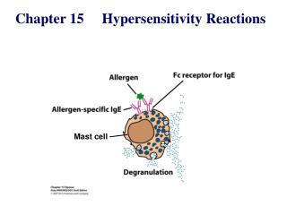
Mast cell degranulation, wheel and flare reaction Ragweed Saline Histamine
MAST CELL RESPONSE TO SURFACE FcRεI CROSSLINKING EARLY MEDIATORS Biogenic amins – histamin Enzymes – triptase, chymase, carboxypeptidase LATE MEDIATORS
The effect of mast cell degranulation varies with the tissue exposed to allergen
Systemic anaphylaxis is caused by allergens that reach the blood stream
Short/Common ragweed (Ambrosia artemisiifolia) Mugwort (Artemisia vulgaris) Green leaf back White leaf back
Mugwort (Artemisia vulgaris) – ? Wormwood (Arthemisia absinthium) – Absinthe (thujone: max 35 mg/l)
HYPERSENSITIVITY REACTIONS INDUCED BY IMMUNE COMPLEXES TYPES II and III • cells expressing the antigen become sensitive to complement mediated lysis or to opsonized phagocytosis • frustrated phagocytosiss tissue demage • the antibody inhibits or stimulates target cell function • no tissue damage (e.g. M. gravis – receptor blocker antibodies) Type II hypersensitivity IgG tpye antibodies bound to the cell surface or to tissue antigens
MECHANISMS OF TYPE II HYPERSENSITIVITY REACTIONS Hemolytic anemia of newborns Erythroblastosis fetalis Drug induced Hemolytic anemia Trombocytopenia Agranulocytosis Penicillin-based antibiotics Anti-arythmic quinidin Goodpasture syndrome (type IV collagen) Pemphigus vulgaris (desmosomal antigens) Damage of epidermal and mucosal junctions, acantholysis
Th B DEVELOPMENT OF DRUG SENSITIVITY Drug-modified cell surface protein Healthy cell IgG type antibodies
FRUSTRATED PHAGOCYTOSIS MEDIATED BY IgG TYPE ANTIBODIES Binding Opsonization Internalization Enzyme release The tissue, which can not be phagocytosed, is damaged Internal or absorbed antigen (drug) Opsonized surface Binding Frustrated Enzyme release phagocytosis
Examples - Type II hypersensitivity Newborn haemolytic anaemia Transfusion reaction Hyperacut allograft rejection Drug-derived • Haemolitic anaemia • Thrombocytopenia • Agranulocitosis • Penicillin-based antibiotics • Anti-arithmic quinidin Goodpasture syndrome (kidney, membrane basalis, collagen type IV) Myasthaenia gravis (anti-acetyl-choline receptor antibodies) Basedow-disease (anti-TSH-receptor antibodies) Pemphigus vulgaris (mucosal bubbles) against desmosomal antigens, interruption of epidermal and mucosal connections, acantolysis (desintegration into single cells)
TYPE III HYPERSENSITIVITY Antibodiesbinding to solubleantigens Small circulating immune complexes Depends on: Size of immune complexes Antigen-antibody ratio Affinity of antibody Isotype of antibody
PATHOGENICITY OF IMMUNE COMPLEXES • Pathogenic immune complexes • Formed in the blood and than are deposited in tissues • Formed in situ at the site of antigen localization • Mechanisms of tissue demage is independent on the site of deposition • Steps of tissue demage • Formation immune complexes in the blood • Deposition depends on the size, composition and cytophylic properties of the antibody (IgM, IgG, IgA) • FcγRIII has a pivotal role – expressed by basophylic granulocytes, NK cells • Permeability of endothelium • Tissue demage • Increased permeability of blood vessels • Reqruitment of neutrophils – enzymes, chemoattractans, dilatators, prostaglandins • fibrosis • Consequences of tissue demage depends on the site of deposition • Arthus reaction – local reaction in skin • Infectious diseases – morbilli – erythema, vasculitis • Acute serum disease – 7 – 10 days • Polyclonal antibodies against snake venom produced in horses (anti-streptococcal) • Immune suppresszive anti-lymphocyte globulin • Bacterial trombolytic streptokinase – treatment of miocardial infarction • Subacute bacterial endocarditis – pathogens are not eliminated • Chronic viral hepatitis • SLE – small vessels, kidney, joints, skin, heart, serosal surfaces
THE PROCESS OF TISSUE DAMAGE CAUSED BY IMMUNE COMPLEXES Blood vessel wall permeability Frustrated phagocytosis Immune complexes activate the complement system, neutrophils, bazophil granulocytes and thrombocytes
Arthus-reaction • Localized Type III hypersensitivity • Local vasculitis develops as a result of immune complex deposition • Inhaled antigens (fungi, animal feces) may induce similar reaction in the lung • IgG type antibody • ‘Farmer lung’ and ‘piegeon breeder lung’
ANA Anti -nuclear antibody
TYPE IV HYPERSENSITIVITY REACTION T CELL MEDIATED PROCESS MACROPHAGES ARE INVOLVED
Type IV hypersensitivity reaction Chemokines, cytokines, cytotoxins
Delayed-type hypersensitivity (DTH) (e.g., tuberculin skin test) TH1 from a previous immunization (memory)
Tuberculin skin test Ag = antigen Mycobacterium protein (PPD) Introduction of Ag
DTH as a result of a contact-sensitizing agent* Contact Dermatitis *a contact-sensitizing agent is usually a small molecule that penetrates the skin then binds to self-proteins, making them “look” foreign
Poison ivy Anacardiaceae (family), Toxicodendron (genus) Toxicodendron radicans or Rhus toxicodendron
Delayed-type Hypersensitivity A positive tuberculin skin test is a DTH reaction
Transplantation reactions • REJECTION • HLA-A, B, C, DR, DQ, DP, minor histocompatibility antigens • foreign MHC-antigens recognized by T cells Direct: self T cells - donor APCs CD8+ T cells Indirect: self APC presents donor MHC-derived peptides CD4+ T cells inflammatory cytokine release • Hyperacut rejection Causes: previous immunization against alloantigens, preformed anti-HLA-antibodies, blood group incompatibility, xenotransplantation antibodies bound to endothel activation of the complement system thrombosis in venules vascularis necrosis Therapy resistent
HLA typing MHC I: HLA-A, HLA-B, HLA-C MHC II: HLA-DP, HLA-DQ, HLA-DR • used for transplantations • Most important is the most polymorphic HLA-B and HLA-DR, HLA-C does not matter) • diagnostical value • Connections between the HLA alleles and diseases Serotyping - microcitotoxicity tests (1) Based on the serological reaction between the TARGET cells and the typing serum. Complement-mediated lysis induced by antibodies recognizing MHC I and/or MHC II cell surface molecules as antigens There is NO reaction in the case of serotype identity (dead cells can be visualized by specific dye) Typing sera containing antibodies to Class I and II proteins are collected from multiparous women, or individuals who had received multiple blood transfusions (immunized against multiple HLA alleles). The specificity of thes polyclonal test sera has been validated by international workshops. The procedure is carried out in Terasaki microtiter plates (with 10µl working aliquots) due to the limited quantity of typing sera. Polyclonal typing sera have been replaced by monoclonal antibodies with unlimited availability.
Serotyping - technology HLA-D (MHC II) antigens are also studied on nylon column separated B cells • The polymorphism of HLA-D antigens could be studied by mixed lymphocyte reactions (MLR) • Bidirectional MLR – lymphocytes of both donors are responding • Unidirectional MLR – lymphocyte proliferation of one donor is blocked (irradiation, Mitomycin treatment) • THE RATIO OF ALLOREACTIVE LYMPHOCYTES IS AN INDIVIDUAL IS 1 – 10 % Microtiter plates
Serotyping have some limits: – crossreactions (HLA-B27 – HLA-B7) – there are no sera available against HLA-C because of its low immunogenicity – some subtypes cannot be discriminated HLA-A 0201 0202 0203 … Genomic DNA based examinations • PCR-SSP is using the PCR amplification reaction directly to detect HLA polymorphisms. Primers can be constructed specifically to complement HLA polymorphisms; if the primers bind the complementary polymorphism and amplify the gene segment, then the PCR product can be detected by standard techniques. It has been constructed an array of PCR primers complementary to the range of HLA polymorphisms