Download
1 / 23
260 likes | 670 Views
Optoelectronics and Measurement Techniques Laboratory. TiO 2 nanoparticles as UV protectors in skin. Doctoral dissertation Alexey Popov. University of Oulu, November 21, 2008. Outline. Solar spectrum UV action spectrum Titanium dioxide: crystal forms Skin structure

E N D
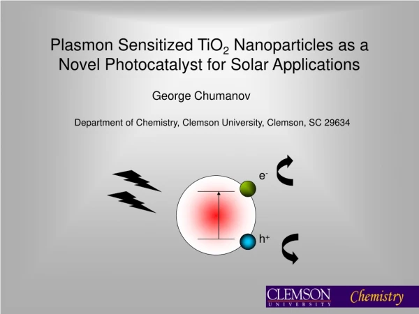
Optoelectronics and Measurement Techniques Laboratory TiO2 nanoparticles as UV protectors in skin Doctoral dissertation Alexey Popov University of Oulu, November 21, 2008
Outline • Solar spectrum • UV action spectrum • Titanium dioxide: crystal forms • Skin structure • Tape stripping technique • TiO2 nanoparticles in horny layer • Calculations by Mie theory • Model of SC with TiO2 nanoparticles • Effect of TiO2 nanoparticles • Comparison with experiment • Conclusion I
EPR setup and samples • Spectrum of sun simulator • TiO2 nanoparticles • EPR measurements • Conclusion II
Solar spectrum Solar spectrum Absorption in stratosphere Wavelength, nm Wavelength, um UV range UVC: 100 – 280 nm (absorbed by ozone layer) UVB: 280 – 315 nm UVA: 315 – 400 nm reach Earth’s surface
UV action spectrum A.P. Popovat al., J. Phys. D: Appl. Phys.38, 2564-2570 (2005).
Titanium dioxide: crystal forms Anatase Rutile Courtesy “Millenium Chemicals”
Skin structure epidermis An OCT image of human skin in vivo (flexor forearm) stratum corneum epidermis dermis Photograph of human corneocytes on a tape strip obtained by Ar+ laser scanning microscopy (λexcit= 488 nm); image size is 250 um x 250 um.
Tape stripping technique Homogeneous distribution Application of the emulsion Pressing of the tape by a roller Removing of the adhesive film J. Lademannat al., J. Biomed. Opt..10, 054015 (2005).
TiO2 nanoparticles in horny layer 0 0 Depth, um 20 0 14 Conc. TiO2particles, ug/cm2 0 In-depth particlesdistribution Volume concentration of TiO2: A.P. Popov et al., J. Opt. Technol.73, 208-211 (2006).
Calculations by Mie theory Opt. properties ofTiO2 particles (rutile modification) Qs =s / (d2)– scattering efficacy factor s– scattering cross-section Qa= a / (d2)– absorption efficacy factor a– absorption cross-section d–particle diameter A.P. Popovet al., J. Biomed. Opt.10, 064037 (2005).
-scat. coef. of nanoparticles - abs. coef. of nanoparticles air - SC phase function - scat. coef. epidermis - abs. coef. hybrid phase function Model of SC with TiO2 nanoparticles Optical parametersfor SC without nanoparticles (adopted from V.V. Tuchin, 1998) Optical parametersfor SC with nanoparticles A = s(1)/(s(1) +sm)
Effect of TiO2 nanoparticles Absorption in the upper part of the horny layer (1-um-thick, with TiO2 particles) (a), reflectance from (b) and transmittance through (c) the whole 20-um-thick horny layer of the incident radiation with = 310 and 400 nm.
Effect of TiO2 nanoparticles The effect of the optimal TiO2 particles (sizes 122 (a) and 62 (b) nm) distributed homogeneously within the 1-um-thick upper part (volume concentration 5%) of the 20-um-thick layer for 400- (a) 310-nm (b) light A.P. Popovet al., J. Biomed. Opt.10, 064037 (2005).
Comparison with experiment Experiment NanoparticlesUV-TITAN M 160 (Kemira, Finland) in absorbing emulsion(L’Oréal, France) Monte Carlo simulations TiO2particles(d = 100 nm,C= 0.2%) intransparent medium (thickness 20 um, nm= 1.4)
Conclusion I Optimal sizes of TiO2 nanoparticles for attenuation of: 310-nm UV light are 62 nm, 400-nm UV light are122 nm. Good correlation with experiment
EPR setup and samples Punch biopsies from porcine ears EPR setup (1.5 GHz) Placebo with PCA and TiO2 (diam. 400 nm, 0, 25 nm) on glass plates, 2 mg/cm2
TiO2 nanoparticles d = 25 nm (a) d = 400 nm (b) TEM photos (TiO2 in emulsion), magnification: x110 (a) and x22 (b). Scale: bar corresponds either to 0.2 um (a) or 1 um (b). Courtesy E.V. Zagainova
TiO2 nanoparticles: anatase Signal of Raman scattering (a); relative absorption efficiency factor (Qa/d) for two wavelengths (b)
EPR measurements EPR signals from placebo with TiO2 particles on glass slides
EPR measurements (a) (b) EPR signals from placebo with 25- (a) and 400-nm (b) TiO2 particles on porcine skin A.P. Popovet al., J. Biomed. Opt.14, xxxxxx (2009).
EPR measurements EPR signals from placebo on porcine skin (a) and skin (b) without particles A.P. Popovet al., J. Biomed. Opt.14, xxxxxx (2009).
Conclusion II If applied onto glass: small particles of 25 nm in diameter produce an increased amount of free radicals compared to the larger ones of 400 nm in diameter and placebo itself. If applied onto porcine skin: there is no statistically distinct difference in the amount of radicals generated by the two kinds of particles on skin and by the skin itself. This proves that: although particles as part of sunscreens produce free radicals, the effect is negligible in comparison to the production of radicals by skin.