Download
1 / 57
570 likes | 598 Views
p53, the most frequently mutated gene in human cancers, serves as the guardian of the genome and tissue, activating pathways in response to DNA damage. Through its distinct domains, such as the DNA-binding domain (DBD), p53 triggers a series of signaling cascades that lead to cell cycle arrest, DNA repair, and apoptosis. Mutations in p53, especially in the DBD, disrupt its functions and can contribute to cancer development. Understanding the activation pathways of p53 is crucial for targeting it as a therapeutic strategy in cancer treatment.

E N D
p53 - guardian or the genome + guardian of the tissuse
p53 • product of a tumour supressor gene • the most frequently mutated gene in human cancers • 393 aa with 4-5 functional domains • biological role as watch dog - “vaktbikkje” • Guardian of the genome - stops the cell cycle upon DNA-damage • Signalling pathway: DNA-damage enhanced p53 activation of CDKI p21 G1 arrest activation of GADD45 stimulated DNA-repair • Guardian of the tissue - facilitates apoptosis if necessary • Signalling pathway: DNA-damage enhanced p53 apoptosis
p53 domains C-terminal allosteric domain
5 distinct domains in p531. TAD • TAD N-terminal [aa1-42] • aa 13-23 conserved between species • F19, L22 and W23 necessary for transactivation in vivo • F19, L22 and W23 involved in binding to TAFII70 and TAFII31 • TAD negatively regulated through interaction with the MDM2 factor or the E1B-55Kd protein • Structure of the MDM2 N-terminal domain + 13-29 peptid from p53 • MDM2 deep hydrophobic pocket • p53 peptide amphipatic helix fitting in the pocket • F19, L22 and W23 involved in binding
Mdm2 - p53 p53-TAD Mdm2
p53 domains C-terminal allosteric domain
5 distinct domains in p532. Pro-rich domain • Et Pro-rich region between TAD and DBD • PxxP present 5 locations in the region 61-94 • deletion of P- rich region reduced apoptosis-response and reduced cell cycle arrest, but normal transcriptional response • contains residues that become phosphorylated upon apoptotic response (HIPK2 phosphorylation of S46)
p53 domains C-terminal allosteric domain
p53 DBD 2 “-helical loops” that contact DNA Zn++ structuring CHCC-Zn++ Two large loops (L2 and L3) involved in minor groove contact Scaffold: -sandwich (two antiparalel -sheets)
5 distinct domains in p533. DBD • DBD centrally located [aa102-292] folded into a “loop-sheet-helix” motif (LSH) • protease-resistent, independent, Zn++-containing domain [CHCC-Zn++] • scaffold: 4- and 5-thread antiparallel -sheet structure • 2 protruding -helical loops contacting DNA directly • Specific base contact in major groove (K120, C277, R280) • Two large loops (L2 + L3) involved in minor groove contact (contact involve R248) • Several H-bond contacts with sugar-phosphate-chain (R273) • Two types of hotspot-mutants in human cancers • disrupts direct DNA-interactions (R248, R273) • disrupts the structure of DBD
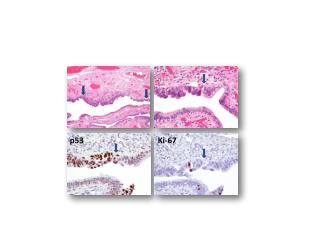
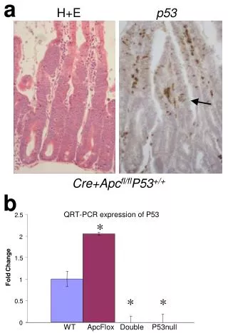
DBD mutations • Most of the p53 mutations that cause cancer are found in the DNA-binding domain • most common mutation changes arginine 248 (red), snaking into the minor groove of the DNA - a strong stabilizing interaction. • Other key sites of mutation are shown in pink, including arginine residues 175, 249, 273 and 282, and glycine 245. Some of these contact the DNA directly, and others are involved in positioning other DNA-binding amino acids.
5 distinct domains in p533. DBD • binds DNA as tetramer (dimer of dimer) • DNA recognition sequence reflects this: 4x RRRCW arranged like this:
p53 domains C-terminal allosteric domain
5 distinct domains in p534. Tetramerization • Tetramerization domain aa 324-355 • 2 + 2 structure • forms tetramers • linked with DBD via 37aa flexible linker [aa 287-323]
p53 domains C-terminal allosteric domain
5 distinct domains in p535. C-terminal allosteric domain • DNA/RNA-binding C-terminal (last 26aa) • open protease-sensitive domain • Basic region • binds DNA and RNA non-specifically and can stimulate annealing • binds DNA ends, internal loops or other loose ends from damaged duplexes • possible function: (allo)steric regulator of specific DNA-binding • p53 appears to be present in a latent form inactive in seq.spec. DNA-binding • Several events in the C-terminal can reactivate p53s central DBD • deletion of basic C-terminal • phosphorylation of S378 with PKC • phosphorylation of S392 with CK2 • binding of C-terminal antibody PAb421 • small singlestranded DNA oligos
. . . . Signal transduction pathways + + p53 + Upstream and downstream p53 functions as sensor of upstream signals reflecting DNA-damage /cellular stress Upstream activation Downstream
Activation of p53 - what happens? • DNA-damage/stress • 1.activation of latent p53 [latent form active form] • enhanced DNA-binding activity • probably also enhanced transactivation activity • post-translational modifications • 2. stabillization and a rapid increase in protein level activation of response • activation level increases 10-100x • Since enhanced levels of p53 may lead to cell cycle-arrest and apoptosis, it is of critical importance that normal cells keep their p53 levels low
Activated by several signals • types of activating stress • DNA-damage (chain breaks, repair-intermediates, recombination-intermediates) • Hypoxia • protective function in tumours (tumour growth limited blood supply hypoxia p53 activation apoptosis of tumour) • trombospondin appears to be p53 regulated, acts antiangiogenic, will reduce blood supply further • NTP pool reduced • sufficient NTP-pool for DNA-replication sensed by p53 • Activated oncogenes (Myc, Ras, E1A, ß-catenin) • Foster defects
The key to stabillization: the MDM2-p53 coupling • MDM2 associates with p53s TAD (aa 17-27) • MDM2-binding leads to • 1. Repression of transactivation • 2. Destabillization of p53 since MDM2-binding stimulates degradation of p53 • mdm2 knock-out = lethal, rescued by simultaneous deletion of p53
The key: MDM2-p53 coupling • Mechanisms for stimulated degradation • MDM2 = p53-specific E3 ubiquitin protein ligase • MDM2 cause transport of p53 from nucleus to cytoplasma, and export is necessary for degradation • MDM2 = a target gene for p53 being activated by p53 • Negative feedback loop - mechanism for turning off the p53 response • Induced relatively late - leaves a time window where p53 can function • regulation = f (MDM2-p53 contact) • Via phosphorylation • Via associated proteins
Several strategies to break theMDM2-p53 coupling Before activation Activated phosphorylation Broken binding Activated phosphorylation inactivated E3-act Activated ARF-binding inactivated E3-act
Recent news • More E3 enzymes suggesting ubiquitylation independent of Mdm2
Regulation of MDM2-p53 contact through phosphorylation of p53 TAD • The ATM kinase • a kinase that is the product of the ATM gene that is lost in pasients with ataxia-telangiectasia • phosphorylates S15 • Weakens the p53-MDM2 interaction • CHK2 - recently identified as a S20-kinase • HIPK2 - recently identified as a S46-kinase • activated as response to UV, role in apoptotic response • DNA PK • DNA-dependent protein kinase • phosphorylates S15 • Weakens p53-MDM2 interaction
upstream signalling pathway Chk2 is a protein kinase that is activated in response to DNA damage and may regulate cell cycle arrest. Chk2-/- cells were defective for p53 stabilization and for induction of p53-dependent transcripts such as p21 in response to gamma irradiation. Chk2 directly phosphorylated p53 on serine 20, which is known to interfere with Mdm2 binding.
Updated: p53 & DNA damage p53 functions as a ‘molecular node’ in the DNA-damage response.
Recent : HIPK2 binds and phosphorylates p53 after UV irradiation UV HIPK2 …leading to apoptosis
Many covalent modifications of p53 in regulatory N- and C-terminal • Phosphorylation • Acetylation • Glycosylation • SUMOylation • Methylation 20
Several modifications - complex regulatory mechanisms • The C terminus of p53 is rich in lysines, which are subjected to acetylation, ubiquitination and sumoylation. • Acetylation of the C terminus • has been shown to protect p53 from ubiquitination. • Acetylation of p53 at K373 and K382 increases its DNA-binding activity and potentiates its interaction with other transcription factors. • The positive effects of acetylation on p53 activity can be reversed by deacetylation. • p53 has also been shown to be sumoylated at K386 • although the exact role of this modification in the regulation of p53 is not yet clear.
p300 Mdm2 Acetylation upon p53 activation p53 stabilization activation • Phosphorylation • followed by • Acetylation 20
Methylation of p53 • A novel mechanism of p53 regulation through lysine methylation by Set9 methyltransferase. • Set9 specifically methylates p53 at one residue within the C-terminal regulatory region. • Methylated p53 is restricted to the nucleus and the modification positively affects its stability. • Set9 regulates the expression of p53 target genes in a manner dependent on the p53-methylation site.
Turning p53 OFF - the hSir2 link • Sir2 - ”silent information regulator” • conserved family identified in silencing in yeast • function as NAD-dep deacetylase
Deacetylation after p53 activation p300 Mdm2 • Phosphorylation • followed by • Acetylation 20 HDAC? = hSIR2
Model DNA damage p53 Stabilized Activated Acetylated Growth arrest Apoptosis Response ON hSir2 Response OFF p53 De-acetylated
N-terminal control via ARF-binding ARF (alternative reading frame) from p16INK4a The INK4A locus (frequently mutated in cancer) →2 alternatively spliced transcripts →translated from alternative reading frames →p16 (cdk-inhibitor) + ARF: •binds MDM2-p53 and inhibits the effect of MDM2s (ligase and shuttling) • ARF strongly induced by viral oncoproteins and contributes to apoptosis of infected cells • ARF also induced by Myc
ARF activation relocalization In unstressed cells, p53 is degraded following interaction with MDM2 and is exported to the cytoplasm using nuclear-export signals present in p53 and MDM2. Inhibition of MDM2-mediated degradation occurs in response to certain stress signals by activation of ARF expression. When ARF binds to MDM2, the MDM2–ARF complex is relocalized to the nucleolus using nucleolar-localization signals present in MDM2 and ARF. This leaves free, transcriptionally active, p53 in the nucleoplasm. Stressed cells ARF expression Unstressed cells p53 degradation Stable p53
Localization: A model for PML-mediated recruitment of p53 to NBs. • PML (promyelocytic leukemia) = org. comp. of nuclear bodies (NBs). • Signal trigger • Signals from DNA damage such as g-irradiation (gIR) or oncogene imbalance – e.g. ras overexpression - trigger SUMOylation (S) and aggregation of PML into NBs. Factors including CBP, Rb, Daxx, Sp100 are also recruited to NBs. • Modification • Consequence: phosphorylation (P) and acetylation (A) of p53. Result in increased and altered p53 transcriptional activity. • Update • PML enhances p53 stability by sequestering Mdm2 to the nucleolus. After DNA damage, PML and Mdm2 accumulate in the nucleolus
DNA damage Oncogene activation Kinase activation ARF activation At least two main pathways Activated p53
Multiple pathways - diverse responses • Multiple pathways exist to stabilize p53 in response to different forms of stress • they may involve down-regulation of MDM2 expression or regulation of the subcellular localization of p53 or MDM2. • Target genes induced by gamma radiation, UV radiation, and the zinc-induced p53 form distinct sets and subsets with a few genes in common to all these treatments.
DNA damage Oncogene activation Kinase activation ? ARF activation ? ? ? ? ? At least two main pathways Activated p53
. . . . Signal transduction pathways + + p53 + Upstream and downstream p53 functions as sensor of upstream signals reflecting DNA-damage /cellular stress Upstream activation Downstream Target genes Activated
p53 as signal transducer- downstream response • downstream consequences leading to repair of damage or apoptosis of damaged cell • Two main types of effects of activated p53 • 1. Stop/regulation of the cell cycle • 2. induction of apoptosis
? Downstream response • Transcriptional effects • Target genes inducing cell cycle arrest • … or DNA damage repair • Target genes promoting apoptosis • Other types of effects?
Cytoplasmic Roles of p53 in Apoptosis • p53 can initiate apoptosis in cells in which trx and translation are inhibited • p53 polyproline domain (aa 62–91) is necessary to cause apoptosis • Excluding p53 from the nucleus causes apoptosis (> threshold level) • p53 affects mitochondrial apoptotic regulators • Cytoplasmically localized p53 can either directly induce Bax oligomerization or liberate proapoptotic BH3-only proteins bound to Bcl2/Bcl-XL at the mitochondria. The released BH3-only proteins can then activate Bax oligomerization and thereby cause cytochrome c release leading to apoptosis.
Regulation of the cell cycle via p53-Rb pathway • Normal cell cycle regulation through four cooperating actors [p16 - cyclin D1 - cdk4 - Rb] which regulate the G1-S transition • most cancers have one of these four altered • p16 negative regulator of cyclin D1/cdk4 • Signalling pathway: DNA-damage activated enhanced p53 activation of CDKI p21 (WAF1, Cip-1) inhibition of cdk4 reduced phosphorylation of Rb G1 arrest • p21 inhibits also several of the other cdks • p21 binds also PCNA stop in replication • Signalling pathway: DNA-damage activated enhanced p53 activation of GADD45 stimulated DNA-repair