Download
1 / 22
220 likes | 345 Views
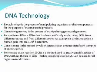
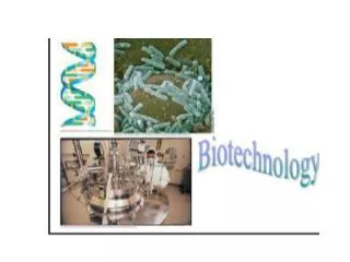
This overview discusses cutting-edge DNA technology, focusing on techniques for copying, analyzing, and recombining DNA. It explains in vitro and in vivo DNA copying methods, gene cloning, and the application of plasmids in gene expression. The role of restriction enzymes in creating recombinant DNA and the significance of sticky ends and DNA ligase in the process are detailed. The document also highlights polymerase chain reaction (PCR) for amplifying DNA segments, gel electrophoresis for DNA separation, and nucleic acid hybridization for specific gene detection.

E N D
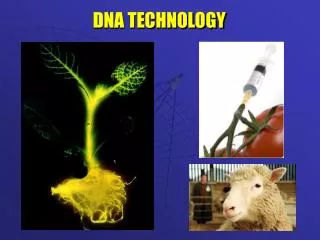
DNA TECHNOLOGY Copying Analyzing Recombining
COPYING DNA DNA can be copied in vitro or in vivo. Genes can be cloned to make a protein of interest or to determine the nucleotide sequence of the gene. The plasmids of bacteria can be used to copy genes.
Recombinant DNA is DNA in which genes from 2 different sources are combined in vitro to make a new molecule. Specific enzyme, called restriction enzymes, (a kind of endonuclease) cut DNA at specific nucleotide sequences. These enzymes come from bacteria. Recognition sequence or restriction site: sequence of nucleotides the enzyme recognizes. Sticky ends:short extensions that CBP with another DNA strand, allows the DNA to be inserted into another genome DNA ligase: seals the 2 strands together
Use of vector to make recombinant DNA Cloning vector: the original plasmid from the bacteria Both the plasmid and the DNA of interest must be cut with the same restriction enzyme Only bacteria with the new gene are grown. This is accomplished by also inserting a gene for antibiotic resistance. Bacteria are cultured on medium that allows for growth of only the transformed bacteria. When the bacteria produce, they make the new gene.
Hybridization with a complementary nucleic acid probe detects a specific DNA within a mixture of DNA molecules. In this example, a collection of bacterial clones (colonies) are screened to identify those carrying a plasmid with a gene of interest. RESULTS APPLICATION TECHNIQUE Cells from each colony known to contain recombinant plasmids (white colonies in Figure 20.4, stap 5) are transferred to separate locations on a new agar plate and allowed to grow into visible colonies. This collection of bacterial colonies is the master plate. Colonies containinggene of interest Master plate Master plate ProbeDNA Solutioncontainingprobe Radioactivesingle-strandedDNA Gene ofinterest Film Single-strandedDNA from cell Filter Filter lifted andflipped over Hybridizationon filter Colonies of cells containing the gene of interest have been identified by nucleic acid hybridization. Cells from colonies tagged with the probe can be grown in large tanks of liquid growth medium. Large amounts of the DNA containing the gene of interest can be isolated from these cultures. By using probes with different nucleotide sequences, the collection of bacterial clones can be screened for different genes. Figure 20.5 The presence of the gene of interest is determined by nucleic acid hybridization- a molecule that has a section of complementary DNA is paired to the gene of interest. The gene can be seen if you use a nucleic acid probe which has a fluorescent or radioactive tag.
Techniques in DNA Analysis: Segments of DNA can also be copied in the lab (in vitro): Polymerase Chain Reaction DNA segments can be separated for analysis: Gel electrophoresis Genomes can be determined by DNA sequencing
3 5 Target sequence APPLICATION With PCR, any specific segment—the target sequence—within a DNA sample can be copied many times (amplified) completely in vitro. 3 Genomic DNA 5 3 5 5 3 TECHNIQUE The starting materials for PCR are double-stranded DNA containing the target nucleotide sequence to be copied, a heat-resistant DNA polymerase, all four nucleotides, and two short, single-stranded DNA molecules that serve as primers. One primer is complementary to one strand at one end of the target sequence; the second is complementary to the other strand at the other end of the sequence. Cycle 1 yields 2 molecules Primers Newnucleo-tides RESULTS During each PCR cycle, the target DNA sequence is doubled. By the end of the third cycle, one-fourth of the molecules correspond exactly to the target sequence, with both strands of the correct length (see white boxes above). After 20 or so cycles, the target sequence molecules outnumber all others by a billionfold or more. Cycle 2 yields 4 molecules Cycle 3 yields 8 molecules; 2 molecules (in white boxes) match target sequence Figure 20.7 Polymerase Chain Reaction ANIMATION
1 2 TECHNIQUE APPLICATION RESULTS Gel electrophoresis is used for separating nucleic acids or proteins that differ in size, electrical charge, or other physical properties. DNA molecules are separated by gel electrophoresis in restriction fragment analysis of both cloned genes (see Figure 20.9) and genomic DNA (see Figure 20.10). When the current is turned on, the negatively charged DNA molecules move toward the positive electrode, with shorter molecules moving faster than longer ones. Bands are shown here in blue, but on an actual gel, DNA bands are not visible until a DNA-binding dye is added. The shortest molecules, having traveled farthest, end up in bands at the bottom of the gel. Mixture of DNA molecules of differ- ent sizes Cathode Each sample, a mixture of DNA molecules, is placed in a separate well near one end of a thin slab of gel. The gel is supported by glass plates, bathed in an aqueous solution, and has electrodes attached to each end. Gel Power source Glassplates Anode Longermolecules Gel electrophoresis separates macromolecules on the basis of their rate of movement through a gel in an electric field. How far a DNA molecule travels while the current is on is inversely proportional to its length. A mixture of DNA molecules, usually fragments produced by restriction enzyme digestion, is separated into “bands”; each band contains thousands of molecules of the same length. Shortermolecules After the current is turned off, a DNA-binding dye is added. This dye fluoresces pink in ultraviolet light, revealing the separated bands to which it binds. In this actual gel, the pink bands correspond to DNA fragments of different lengths separated by electrophoresis. If all the samples were initially cut with the same restriction enzyme, then the different band patterns indicate that they came from different sources. Figure 20.8 Gel electrophoresis
DdeI DdeI DdeI DdeI Normal -globin allele 201 bp Large fragment 175 bp Sickle-cell mutant -globin allele Large fragment 376 bp DdeI DdeI DdeI (a) DdeIrestriction sites in normal and sickle-cell alleles of -globin gene. Sickle-cellallele Normalallele Largefragment 376 bp 201 bp175 bp (b) Electrophoresis of restriction fragments from normal and sickle-cell alleles. Figure 20.9a, b Gel electrophoresis used to isolate alleles of hemoglobin gene
APPLICATION Researchers can detect specific nucleotide sequences within a DNA sample with this method. In particular, Southern blotting is useful for comparing the restriction fragments produced from different samples of genomic DNA. TECHNIQUE In this example, we compare genomic DNA samples from three individuals: a homozygote for the normal -globin allele (I), a homozygote for the mutant sickle-cell allele (II), and a heterozygote (III). Heavyweight Nitrocellulose paper (blot) Restriction fragments DNA + restriction enzyme I II III Gel Sponge Papertowels I Normal -globin allele Alkalinesolution II Sickle-cell allele III Heterozygote 3 2 Blotting. 1 Gel electrophoresis. Preparation of restriction fragments. Figure 20.10 animation
RESULTS Probe hydrogen- bonds to fragments containing normal or mutant -globin I II III I II III Radioactively labeled probe for -globin gene is added to solution in a plastic bag Fragment from sickle-cell -globin allele Film over paper blot Fragment from normal -globin allele Paper blot 1 2 Hybridization with radioactive probe. Autoradiography. Because the band patterns for the three samples are clearly different, this method can be used to identify heterozygous carriers of the sickle-cell allele (III), as well as those with the disease, who have two mutant alleles (II), and unaffected individuals, who have two normal alleles (I). The band patterns for samples I and II resemble those observed for the purified normal and mutant alleles, respectively, seen in Figure 20.9b. The band pattern for the sample from the heterozygote (III) is a combination of the patterns for the two homozygotes (I and II).
Example of gel electrophoresis used to identify an unknown variety of plant.
Restriction Fragment Length Differences as Genetic Markers • Restriction fragment length polymorphisms (RFLPs) Differences in DNA sequences on homologous chromosomes that result in restriction fragments of different lengths • Specific fragments can be detected and analyzed by Southern blotting • The thousands of RFLPs present throughout eukaryotic DNA can serve as genetic markers, can identify genes that cause disease • The Human Genome Project Sequenced the human genome • Scientists have also sequenced genomes of other organisms
Chromosome bands Cytogenetic map Chromosome banding pattern and location of specific genes by fluorescence in situ hybridization (FISH) Genetic markers Genes located by FISH 1 Genetic (linkage) mappingOrdering of genetic markers such as RFLPs, simple sequence DNA, and other polymorphisms (about 200 per chromosome) 2 Physical mapping Ordering of large over- lapping fragments cloned in YAC and BAC vectors, followed by ordering of smaller fragments cloned in phage and plasmid vectors Overlappingfragments 3 3 DNA sequencing Determination of nucleotide sequence of each small fragment and assembly of the partial sequences into the com- plete genome sequence …GACTTCATCGGTATCGAACT… Figure 20.11 Mapping a large genome Construct a linkage map of several thousand genetic markers spaced throughout each of the chromosomes Cut fragments with restriction enzymes, separate with gel electrophoresis 3. Sequence each small fragment animation
RFLP marker DNA Disease-causing allele Restriction sites Normal allele Figure 20.15 Applications of DNA Technology Diagnosis of Disease: bacteria and viruses can be identified from their DNA. Genetic disorders can be diagnosed, carriers can be identified.
Cloned gene (normal allele, absent from patient’s cells) Viral RNA 2 Retrovirus capsid Bone marrow cell from patient Figure 20.16 2. Human Gene Therapy: altering the genome of an individuals DNA. In this case a virus is used as the vector. This works for diseases with one defective gene. Need to change stem cells from bone marrow. So far, this isn’t working too well.
3. Pharmaceuticals Gene that codes for the protein that you want, (like human growth hormone, insulin, vaccine) is transferred into a bacterium or yeast. A highly active promoter is also inserted Host makes large quantities of the protein Protein is purified
Blood from defendant’s clothes Victim’s blood (V) Defendant’s blood (D) 4 g 8 g V Jeans D shirt 4. Forensics DNA fingerprinting to identify a suspect
Figure 20.18 5. Animal Husbandry
USE • Transgenic animals: inserting a gene to produce a trait like more wool, leaner pig, etc. • Inserting a gene for the production of some protein like blood clotting factor in cow milk: “pharm animal” TECHNIQUE • Remove egg from female and fertilize in vitro • Clone the desired gene from another organism • Inject DNA into the nuclei of the egg • Implant egg with new gene into a surrogate mother
6. Genetic Engineering in Plants USE Insert a gene to improve the crop, like delay ripening, resistance to disease. Insert gene to make a desired protein, like Vitamin A in sugar cane TECHNIQUE 1. Vector is a plasmid from a bacteria that causes tumors in plants (crown galls). This technique only works in dicots, other techniques like electroporation and DNA guns are used for monocots like corn, rice and wheat
2 3 1 Agrobacterium tumefaciens Genes conferring useful traits, such as pest resistance, herbicide resistance, delayed ripening, and increased nutritional value, can be transferred from one plant variety or species to another using the Ti plasmid as a vector. APPLICATION RESULTS TECHNIQUE Tiplasmid Site where restriction enzyme cuts The Ti plasmid is isolated from the bacterium Agrobacterium tumefaciens. The segment of the plasmid that integrates into the genome of host cells is called T DNA. T DNA DNA with the gene of interest Recombinant Ti plasmid Isolated plasmids and foreign DNA containing a gene of interest are incubated with a restriction enzyme that cuts in the middle of T DNA. After base pairing occurs between the sticky ends of the plasmids and foreign DNA fragments, DNA ligase is added. Some of the resulting stable recombinant plasmids contain the gene of interest. Recombinant plasmids can be introduced into cultured plant cells by electroporation. Or plasmids can be returned to Agrobacterium, which is then applied as a liquid suspension to the leaves of susceptible plants, infecting them. Once a plasmid is taken into a plant cell, its T DNA integrates into the cell‘s chromosomal DNA. Transformed cells carrying the transgene of interest can regenerate complete plants that exhibit the new trait conferred by the transgene. Plant with new trait Figure 20.19 Plants