Virus Entomopathogens: Structure, Life Cycle, and Applications
740 likes | 1.01k Views
Discover the origins, structure, and life cycle of virus entomopathogens, with insights into their role in controlling insect pests and the advancements in genetic research in this field. Learn how these viruses infect invertebrates and the significance of Baculovirus in biopesticides.

Virus Entomopathogens: Structure, Life Cycle, and Applications
E N D
Presentation Transcript
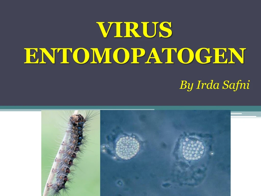
VIRUS ENTOMOPATOGEN By Irda Safni
“Virus” berasal dari bahasa Latin, yang artinya racun dan selalu berhubungan dengan penyakit dan kematian. • Definisi virus (modern): • biosistem paling dasar yang menunjukkan keragaman makromolekuler yang mampu berkembang biak dan membelah sendiri, tapi kurang mampu bereaksi terhadap faktor lingkungan, tidak dapat bergerak sendiri, dan tidak dapat menggunakan sumber energi metaboliknya sendiri bertolak belakang dengan definisi makhluk hidup
Kebanyakan biosistem dasar yang menunjukkan kompleksitas makromolekul dan mampu bereplikasi sendiri dan berevolusi, tetapi kekurangan iritabilitas (kemampuan untuk bereaksi terhadap faktor lingkungan), pergerakan sendiri, dan sumber energi metabolik sendiri, berlawanan dengan sifat dasar yang merupakan ciri-ciri makhluk hidup.
Setelah virus partikel masuk ke dalam sel yang peka, asam nukleatnya akan mengambil alih sistem metabolik sel dan bereplikasi menjadi virus-virus partikel yang baru, hingga isi sel inangnya habis dan akhirnya mati.
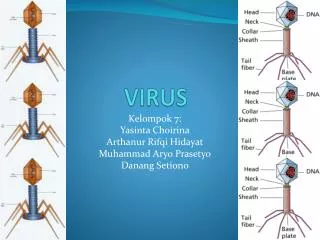
Struktur Virus 1. Mengandung DNA atau RNA 2. Mengandung protein coat (capsid) 3. Reseptor – pada capsid yang menentukan sel inang apa yang dapat diinfeksi dan bagaimana virus menginfeksi sel.
Struktur Virus • Inner core nucleic acid • Mengandung bahan genetik (DNA atau RNA) • Outer core nucleic acid • Mengelilingi virus (Capsid) • Mengandung reseptor
Struktur yang tidak dijumpai pada semua virus • Amplop • mengelilingi beberapa virus hewan • Terbuat dari membran inang • Tail, end plate, tail fibres • Hanya pada Bacteriophage
Siklus Hidup Virus • Virus adalah parasitobligat intracellular, yang artinyamereka tidak dapatbereplikasi atau mengekspresikan gennya tanpa bantuan sel hidup. • Setiap virus memiliki kisaran inang, sejumlah sel inang yang dapat diinfeksinya.
Tahap Perbanyakan Virus • Setelah virus menginfeksi inangnya, komponen turunan virus dihasilkan oleh mesin sel inangnya • Perakitan kapsid virus adalah proses non-enzimatik. Biasanya secara spontan. • Biasanya virus hanya dapat menginfeksi sejumlah kecil inang (dikenal dengan nama "kisaran inang"). • Mekanisme "gembok dan kunci" adalah prinsip utama kisaran inang virus. • Protein tertentu pada partikel virus harus cocok pada tempat reseptor tertentu pada permukaan sel inang.
Siklus Hidup Virus • Untuk dapat bertahan hidup, virus harus mampu melakukan sebagai berikut: • 1. Menemukan sel inang yang di dalamnya virus dapat bereplikasi • 2. Mengikat pada sel • 3. Memasuki sel • 4. Melepas genomnya agar dapat bereplikasi • 5. Mereplikasi genomnya • 6. Transkripsi dan translasi protein virusnya • 7. Membungkus genom dan proteinnya • 8. keluar dari sel
2 1 3 4 Figure 19.4 VIRUS Entry anduncoating DNA Transcriptionand manufacture ofcapsid proteins Capsid HOSTCELL Replication Viral DNA mRNA ViralDNA Capsidproteins Self-assembly ofnew virus particlesand their exit fromthe cell
Penyakit yang disebabkan virus entomopatogen mulai diketahui sejak abad ke-16. • Penyakit yang disebut Jaundice o graserrie, sekarang diidentifikasi sebagai nucleopolyhedrosis, ditemukan pada ulat sutra (Bobyx mory) oleh Vida pada tahun 1524 dan kemudian juga diisolasi dari lebah madu (Apis mellifera). • Pada tahun 1856, dua orang ahli Italia (Maestri dan Cornalia) menjelaskan occlusion bodies (OBs) ulat sutra nucleopolyhedrosis. • Pada tahun 1926 Paillot mendeskripsikan granulovirus (GVs) pertama sekali. • Pada tahun 1934 Ishimori menjelaskan jenis baru polyhedrosis di dalam ulat sutra OBs dibentuk didalam sitoplasma sel yang diinfeksi (bukan pada asam nukleat) sekarang dikenal dengan cypovirus.
Sejak tahun 1950 s/d 1970, Steinhaus dan koleganya menguji Baculovirus sebagai agens hayati di lapangan dengan mengaplikasi nucleopolyhedrovirus (NPV) untuk mengendalikan ulat alfalfa (Colias eurytheme Boisduval; Lepidoptera: Pieridae). • Bioinsektisida komersil berbahan aktif virus pertama dikembangkan pertama sekali pada tahun 1975 oleh Perusahaan Sandoz (dengan nama dagang Elcar) untuk mengendalikan Heliothis/Helicoverpa Lepidoptera: Noctuidae). • Selama tahun 1979 s/d 1980, penemuan penting pada genetika virus entomopatogen, khususnya Baculovirus. • Hingga saat ini studi genetika virus entomopatogen difokuskan pada studi genom lengkap telah ada 29 sekuensing genom lengkap virus entomopatogen.
Sama seperti bakteri, virus entomopatogen harus ditelan oleh serangga inangnya, sehingga efektif untuk mengendalikan serangga yang memiliki tipe mulut pengunyah. • Beberapa serangga Lepidoptera adalah inang penting Baculovirus, termasuk nucleopolyhedroviruses (NPV) dan Granuloviruses (GV).
Virus yang Menginfeksi Invertebrata • Virus DNA • Double stranded DNA • - Poxviridae • - Iridoviridae • - Baculoviridae: NPV & GV • - Polydnaviridae • Single stranded DNA • - Parvoviridae • Virus RNA • Double stranded RNA • - Rheoviridae: Cypovirus • Single stranded RNA (-) • - Rhabdoviridae • - Bunyaviridae • Single stranded RNA (+) • - Picornaviridae, Togaviridae, • Tetraviridae, Flaviridiae, • Nodaviridae
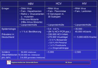
Tabel 1. Kelompok Virus Entomopatogen s ds= double-stranded, ss=single-stranded.
Klasifikasi Virus Entomopatogen • Klasifikasi virus entomopatogen dibuat sesuai dengan peraturan the International Commitee on Taxonomy of Viruses (ICTV). • Virus entomopatogen diklasifikasikan menjadi 12 famili. • Kriteria untuk menyusun klasifikasi disusun berdasarkan keragaman virus serangga, seperti: • Jenis bahan genetik (seperti single- ataudouble-stranded DNA, single- ataudouble-stranded RNA, positive ataunegative strand) • Morfologi dan ukuran virion (seperti icosahedral, rod-shaped, dll.) • Kehadiran amplop di sekeliling virion. • Kehadiran occlusion body (OB) melingkupi virion. • Inang dan kisaran inang.
Klasifikasi Virus Entomopatogen • Kriteria utama sekuensing bahan genetik virus serangga bukan hanya menentukan diskriminasi antar spesies virus, akan tetapi juga membangun hubungan evolusioner antar virus dalam kelompok yang sama.
Penamaan Virus Entomopatogen • Virus entomopatogen diberi nama dengan cara singkatan (akronim). • Sesuai dengan inangnya, dan kelompok virusnya • Contoh: Autographacalifornicamultiple nucleopolyhedrovirusAcMNPV • Semua nucleopolyhedroviruses dinamai NPV, granuloviruses dinamai GV, entomopoxviruses: EPV, iridoviruses: IV, dan cytoplasmic polyhedrosis viruses (cypoviruses) :CPV. • Virus entomopatogen sangat tinggi keragamannya, tapi hanya beberapa kelompok saja yang dijumpai pada populasi serangga dan menunjukkan potensi sebagai agen pengendali hayati.
Life Cycle of Insect Virus Generalized life-cycle of insect viruses. Figure credit: Jim McNeil, Department of Entomology, Penn State University.
Virus particles are usually found on the surface of plants or in the soil. • Insects become infected by consuming plant material with viral particles on the surface, although some pests of low-growing plants can be infected by contact with the soil. • Virus infection begins in the insect’s digestive system but spreads throughout the whole body of the host in fatal infections. • The body tissues of virus-killed insects are almost completely converted into virus particles. • The digestive system is among the last internal organ system to be destroyed, so the insects usually continue to feed until they die. Infected insects look normal until just prior to death, when they tend to darken in color and behave sluggishly. • They often develop more slowly than uninfected individuals.
Most virus-infected insects die attached to the plant on which they feed. • Virus-killed insects break open and spill virus particles into the environment.These particles can infect new insect hosts. • Because of the destruction of the internal tissues, dead insects often have a “melted” appearance. • The contents of a dead insect can range from milky-white to dark brown or black. • While natural virus outbreaks tend to be localized, virus particles can be spread by the movement of infected insects, the movement of predators such as other insects or birds that come into contact with infected insects, or non-biological factors like water run-off, rain-splash or air-borne soil particles.
Many virus-infected insects also climb to higher positions on their host plant before they die, which maximizes the spread of virus particles after the insect dies and disintegrates. • The number of virus infection cycles within a growing season depends heavily on the insect’s life cycle. • Insect pests with multiple generations per season or longer life cycles can be more heavily impacted by virus outbreaks since there is a greater opportunity for multiple virus infection cycles within a growing season.
Advantages and Disadvantages of Insect Viruses for Controlling Pests Advantages • Insect viruses are unable to infect mammals, including humans, which makes them very safe to handle. Most insect viruses are relatively specific, so the risk of non-target effects on beneficial insects is very low. • Many viruses occur naturally and may already be present in the environment. Even in cases where they are applied, successful infections can perpetuate the disease outbreak making repeat applications within a season unnecessary.
Disadvantages • Most insect viruses take several days to kill their host insect, during which the pest is still causing damage. Insect death is also dose dependent, and very high doses are often necessary for adequate control. As insects age, they can become less susceptible to virus infection, so viruses are usually only effective against early larval life stages. • Although viruses can persist in the environment for months or years, exposed virus particles, like those on the surface of plants, are quickly inactivated by direct sunlight or high temperatures, which can limit their persistence within a given season. Also, some agricultural practices can reduce persistence between seasons, such as tillage, which buries virus particles in the soil.
Suggestions for application of insect viruses • Viruses are usually not “stand alone” solutions to an insect pest problem, but are most effective in conjunction with other management strategies. • Insect viruses are fairly specific, be sure that the target pest is correctly identified. • Carefully scout fields before application and apply virus when the target pests are young but actively feeding.
Apply virus to maximize the longevity and effectiveness of virus particles: • Thoroughly coat plants to maximize coverage. Young plants can even be dipped in a solution of virus particles to completely cover the leaf area. • Apply in the morning or evening or on cloudy days when degradation from sunlight is reduced. • Avoid applying on rainy days, as rain will wash virus particles off the leaf surfaces. • Use formulations with ultraviolet (UV) light blockers and sticking agents to increase longevity. • Using mixed cropping and reduce soil disturbance after application. These help increase the persistence of virus particles in the system and may lead to better control within and between growing seasons.
6 Baculoviruses Spodopteralittoralis 2microns FromHunter-Fujitaetal
Kelompok Baculovirus yang penting sebagai virus entomopatogen adalah nucleopolyhedroviruses (NPV) and granuloviruses (GV). • Virus ini memiliki jenis inclusion bodies yang berbeda-beda, di mana virus partikel (virion) menempel. • Parikel virus menyerang inti sel usus kecil, lemak tubuh atau sel-sel jaringan lainnya dan cairan tubuh serangga yang sudah mati. • Sebelum mati, larva yang terinfeksi memanjat bagian yang lebih tinggi dari tanaman, yang membantu diseminasi parikel virus dari tubuh larva yang sudah mati ke bagian tanaman yang lebih rendah. • Kebiasaan ini membantu menyebarkan virus untuk menginfeksi larva yag sehat.
Virus sangat spesifik inang dan dapat menyebabkan pengurangan populasi inang secara nyata. • Contoh virus yang dijual secara komersil: • - Helicoverpa zea single-enveloped nucleopolyhedrovirus (HzSNVP) • - Spodoptera exigua multi-enveloped nucleopolyhedrovirus (SeMNPV) • - Cydia pomonella granulovirus (CpGV).
7 Cara Kerja Baculovirus Baculoviruses- Modeofaction FromHunter-Fujitaetal
8 Baculoviruses SusceptibilityofAlternativeHosts Foundonlyininvertebrates Nomemberofthefamilyisknowntoinfectplantor vertebrate Mosthavenarrowhostinsectrange,andinfectivityis restrictedtotheoriginalhostgenusorfamily
9 Baculoviruses Toxicitystudies-mammals Toxicitytestresultsfrom1970s/80sof29NPVs indicatednotoxicityorpathogenicity.Doseswere generally10–100xthe“peracre”(1acre=0.45ha) fieldrateequatedtoa70kgperson. HeliothiszeaNPVmostextensivelytestedfortoxicityin humansandledtoregistrationof“Elcar”bySandozin USA.
10 Baculoviruses Toxicitystudies–mammalscont. NoeffectsofHzNPVfoundin: Acutetoxicity-pathogenicitytestsinmouse,rat,guineapig,rabbit, monkeyandmanat6x109–3x10¹²OB/kg. Skinirritationsensitivitytestsinguineapigs,rabbitsandmanat106 and107OB/mm2skin. Eyeirritationtestsinrabbitswith105and2x106OB/eye Subacutetoxicity-pathogenicitytestsandsubcutaneousinjectioninto mice,rats,dogsandrhesusmonkeys. Teratogenicityandcarcenogenicitystudiesinratsandmiceat109– 3.5x10¹²OB/kg. SimilarbutlessextensiveresultsformanyotherNPVsfromthe 1970s/80s
11 Baculoviruses Toxicitystudies–wildlife Birds AbletopassNPVthroughthealimentarytractunaffected Nodeleteriouseffects Aquaticorganisms Noadverseeffects Beneficialinsects Nodirecteffectonparasitoids,predatorsandpollinators Indirecteffectsonparasitoidsresultingfromhostdeath
12 Baculoviruses Pathologystudies Toxicitytestsdesignedfortestingeffectsofchemicalson vertebratesareinsufficient ResultsreportedinGröner(1986)indicatenovirusinduced antibodyproductionintestmammalsandchicken. Nocytogeneticeffectsofbaculovirusesinmammaliancellseither invivoorinvitro.
13 Baculoviruses Virus-cellinteractionsinvitro AcNPVinoculatedintovertebratecellscanbetaken- upandthedegreeofup-takedependsoncelltype, temperature,timeandviralphenotype. BUT,noneofthehumanandnonhumanvertebrate linestestedshowedevidenceofviralreplication. NPVsunabletoactivateretrovirusesinmammalian celllines
15 Baculoviruses alistofthebaculovirusesregulatedaspesticide activeingredientsbytheUSEPAOfficeofPesticide ProgramsasofMay2005 AnagraphafalciferaNPV CydiapomonellaGV DouglasfirtussockmothNPV GypsymothNPV HelicoverpazeaNPV IndianmealmothGV MamestraconfigurataNPV(pending) SpodopteraexiguaNPV
16 Baculoviruses–USEPAfactsheet III.ASSESSINGRISKSTOHUMANHEALTH Thesevirusesinfectonlythetargetinsectlarvaeandcloselyrelated species.Toxicitytestsshowthatthevirusesposenorisktothepublic. Workerswearprotectiveclothingtopreventpossibleirritationfrom handlingandapplyingtheproduct. IV.ASSESSINGRISKSTOTHEENVIRONMENT TestsshowthattheGVandNPVsthatEPAhasregisteredaspesticide activeingredientsspecificallyinfectonlycertainspeciesofmothlarvae. Thevirusesdonotharmotherorganisms,includingplants,beneficial insects,otherwildlife,ortheenvironment.Thesevirusesoccurnaturally intheirinsecthosts.
17 Cypoviruses:Modeofaction Polyhedraingestedanddissolvedinlarvalmidgut Virionsreleasedandattachtomidgutcolumnarcells Viralcoreenterscellcytoplasm RNAtranscriptionandreplication RNAoccludedincapsules Viruscapsulesoccludedbyvirogenicstromatoformocclusion bodies
18 Cypoviruses(Rheoviridae) NoCPVhasbeenfoundinfectingvertebratesorplants (Belloncik,1989) DendrolimusspectabilisCPVregisteredinJapanin 1974.Safetytestresultsgenerallynegative. –Katagiri,K.(1981)Pestcontrolbycytoplasmicpolyhedrososviruses.In: Microbialcontrolofpestsandplantdiseases1970-1980.(EdBurges,H.D.) AcademicPress.
Poxviridae:Entomopoxviruses NPV GV NPV GV
Member of the family of Poxviridae has a wide host, including vertebrates and invertebrates. • Chicken pox and Small pox virus belong to this family. • The show allantoid – to brick-shaped virions, occluded within ovoid OBs called Spheroids. • Entomopoxvirus has been isolated from 27 orthopterans, lepidopterans, dipterans and coleopterans. • The subfamily Poxvirinae includes three genera, i.e. EntomopoxvirusA, EntomopoxvirusB, and EntomopoxvirusC • Entomopoxvirus A infects only coleopteran species; EntomopoxvirusB infects lepidopteran and coleopteran species; Entomopoxvirus C infects only dipteran species. • The fourth group, group D, has been proposed by ICTV which only attacks hymenopterans.
Poxviridae:Entomopoxvirus Anomala cuprea (Coleoptera) larvae infected with an entomopoxvirus show the symptoms of the infection such as a whitish appearance and underdevelopment (left, infected larva; right, healthy one).
Poxviridae:Entomopoxvirus FIG. 1. Material from Anomala cuprea larvae used for real-time quantitative PCR. (A) Dissected midgut consisting of most of the whole midgutof a first-day second-instar larva (the PM inside has been removed). There is a circle of prominent sac-shaped structures (referred to here as“sac-circles”) on both the anterior and the posterior regions of the dissected midgut. Those on the anterior region of the midgut may be ceca. Thesetwo circles with portions of midgut were cut away from the rest of the midgut with a sharp edge (incision lines are indicated in the picture) andwere combined into a single sample. (B) Separated sac-circles with a portion of midgut from the rest of the anterior region of the midgut ofsecond-instar larva. Each sac contains digestive juices that passed through the PM from the endoperitrophic to the ectoperitrophic space (viewed from an angle different from that in panel A). (C) Diagram of a second-instar A. cuprea larva. Part of the integument has been omitted so thatthe materials are visible.