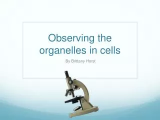
Observing Cells
Do Now: Your digestive system uses many different enzymes to digest food . Pepsin is an enzyme in your body that breaks down proteins in food that you eat. 1. What is pepsin made of? 2. What is pepsin’s substrate?

Observing Cells
E N D
Presentation Transcript
Do Now: Your digestive system uses many different enzymes to digest food . Pepsin is an enzyme in your body that breaks down proteins in food that you eat. 1. What is pepsin made of? 2. What is pepsin’s substrate? 3. Pepsin is found in the stomach. Would you expect it to work best at low, medium, or high pH? 4. Your body temperature is about 37oC. What will happen to pepsin if it is used at 20oC in a lab? Observing Cells
Lab Tomorrow: Observing Cells • Tomorrow’s Lab will have 3 parts: • Figuring out the field of view size • Observing a live plant cell • Observing a prepared animal cell slide
Microscope Use • FIRST: make sure the light is on, and the LOW POWER objective lens (RED) is in place before doing anything. • Place the slide on the stage. • Use the big knob (coarse focus) to adjust the focus
Medium & High Power • NEVER USE THE BIG KNOB TO FOCUS ON MEDIUM OR HIGH POWER. • You might break the slide or lens! • Some of our microscope’s high power lenses aren’t very clear. My apologies if you get one, but the lab will still work just fine.
Lenses • Every lens has a magnification power. A 10x lens, for example, magnifies things to 10 times actual size. • Compound microscopes like the ones you will use have 2 lenses: the eyepiece, and an objective lens.
Magnification • The total magnification is found by multiplying the power of lenses together. • For example, a 10x eyepiece used with a 50x objective gives a total magnification of (10)(50) = 500x • This will help you complete table 1. • Eyepiece = 10x • Low = 4x • Medium = 10x • Hi = 40x
Part 1: Field of View Size • You will measure the field of view size on LOW POWER using a clear ruler. • Measure to the nearest 0.1 mm (estimate) • You will then use the formulas given on your lab handout to calculate the medium & hi power FOV sizes.
Part 2: Animal Cell Observation • You will observe animal cells on low, medium, and hi power (if available) • You will sketch what you see on medium power (you may do hi if it works, but change the title if you do) • Label the cell’s nucleus & cell membrane • Describe the cell’s shape, etc. • Be accurate! Draw cells approximately the same size and shape as they appear!
Part 3: Plant Cell Observation • You will make a plant cell wet-mount slide • Use forceps to pluck a single leaf of elodea. • Place it on the slide, add 1-2 drops of water • Place a cover slip on the slide, and put it on the stage • Observe the plant cell as you did the animal cell • Label a cell wall and a chloroplast
Determining Cell Size • You will use your observations to calculate the sizes of the cells you observe. • First, figure out the medium power FOV size • Divide that number by how many cells fit across the FOV • Multiply by 1,000 to convert mm to µm
Discussion Questions • There are 4 discussion questions for you to answer after completing the lab.