Download
1 / 49
500 likes | 773 Views
WHOLE-BODY-LOW-DOSE MDCT IN THE INVESTIGATION OF MULTIPLE MYELOMA (MM) – A NEW APPROCH AND OUR EXPERIENCE Kamenetsky Natalya (1), Rachmilewitz Eliezer (2), Katz Rama (1), (1)Department of Diagnostic Imaging (2) Department of Heamatology E. Wolfson Medical Center, Holon, Israel.

E N D
WHOLE-BODY-LOW-DOSE MDCT IN THE INVESTIGATION OF MULTIPLE MYELOMA (MM) – A NEW APPROCH AND OUR EXPERIENCE Kamenetsky Natalya (1), RachmilewitzEliezer (2), Katz Rama (1), (1)Department of Diagnostic Imaging (2) Department of Heamatology E. Wolfson Medical Center, Holon, Israel.
The idea of our study came from lately published literature, especially the article: “Whole-body low dose multidetector row-CT in the diagnosis of MM: an alternative to conventional radiography” EJR,2005.
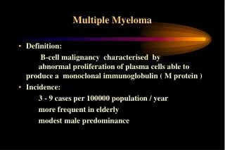
MM – Definition and diagnosis Uncontrolled proliferation of neoplasticplasma cell clone in the bone marrow. Diagnosis based on laboratory and radiographic findings: • Bone marrow containing more then 15% plasma cells (normally no more then 4%). • Blood serum or urine containing an abnormal protein (M protein, Bence-Jones protein). • Bone lesions found on skeletal survey as generalized osteopenia or lytic bone deposits.
MM – Demographics: • Most common primary bone tumor in adult. • Multifocal lesions more common • Solitary (Plasmacytoma) less common: may be Intra/Extraosseous. • Age: 40 years or older. • M:F = 2:1 • More common in Afro-Americans then in Caucasians. Less common in Asians. • Median survival: 3-4 years.
MM – Skeletal involvement: Osteolytic lesion(80%) - found particularly with nodular marrow infiltration. small discrete lytic areas of bone destruction with no reactive bone formation. Arises within the medulla, may progress to infiltrate the cortex and periosteum and be accompanied with extraosseous soft tissue masses.
MM-Skeletal involvment: • Diffuse osteopenia(85%) is associated with a packed pattern of marrow infiltration – thinning of all trabeculae, vertebral body collapse. • Osteosclerosis – rare (1-3%), may be focal or diffuse. • Normal survey (10%).
Skeletal involvement in MM: Frequency in different bones correlates with normal sites of red marrow distribution : • Vertebra (66%). • Ribs (45%). • Skull (40%). • Shoulder (40%). • Pelvis (30%). • Long bones (25%).
CT versus plain film: • Bone lesions of the axial skeleton, are significantly better recognized byCTby reducing the effects of overlying soft tissue and bony structures. • Bone lesions of the appendicular skeleton are mostly well recognized in both modalities.
Roll of imaging in MM patients: • Diagnosis and staging. • Diagnosis of extramedullary or solitary plasmacytoma and directing a biopsy if needed. • Monitoring treatment response. • Detection of relapse. • Assessing fracture risk and directing prophylactic treatment.
Staging by Durie and Saimon: Stage 1:Stage 3: (All) (1 or more) Hemoglobin >10g/100ml <8.5 g/100ml Serum calcium <12mg/100ml >12mg/100ml M component IgG: <5g/100ml >7g/100ml IgA:<3g/100ml >5g/100ml Urine light chain <4 g/24hr >12g/24hr Bone Lesionnone /solitarymultiple Stage 2: Between Stage 1 and 3.
MM – Staging: Patients with more then two unequivocal lytic lesions are classified as stage 3, indicating immediate treatment.
Different imaging modalities in MM: X-ray – Conventional plain film survey, CT. MRI Radionuclid imaging – Tc(99m)- MIBI, F-18 FDG-PET.
Plain film skeletal survey: Multiple lytic lesions (80%). Solitary (Plasmacytoma) expansible lytic lesion. Osteopenia (85%). Vertebral body collapse and pathological fractures. Normal survey (10%). Shrinking or sclerosing deposits indicate a response. Residual osteolysis may persist in inactive phase of disease. No detection of extraosseous involvement.
CT Imaging: • Detect disease in bone, bone marrow and extramedullary sites. • Focal pattern – sharp, lytic lesions with no sclerotic rim. • Diffuse faint osteolysis. • High (soft tissue) attenuation value of bone marrow. • Positive response to treatment – Shrinking or sclerosing deposit, disappearance of soft tissue masses, reappearance of cortical contour and fatty marrow content.
Our experience: • On April - November 2006 we performed 41 CT skeletal surveys: • 30 patients with known diagnosis of MM. • 5 to exclude MM lesion in MGUS patients. • 6 in other patients.
CT survey study protocol: • Patient laying supine, cranio-caudal position, arms on abdomen. • Scan length from top of the skull down to the end of the knees. • With suspended respiration when possible. • No oral or IV contrast material.
CT survey study protocol:* Low dose CT parameters are based on the article from EJR 2005. • MDCT 16 slices. • Surview 1536 mm. • 120 KV, 70 mAs (300 mAs in spine CT) • Overall radiation dose of 5 mSv. • 16*0.75mm collimation with 0.5 sec rotation time. • Table speed – 18mm/sec. • Slice thickness – 3mm. • Mean acquisition time – 38 sec.
CT survey study protocol: • Reconstruction was done from raw data. • bone filter with B60f kernel. • F.O.V = 500mm max. • multiplanar reformatted (MPR) whole body images were reconstructed in sagital and coronal planes. Divided into 3 different body parts: • Head and neck, including cervical spine • Chest and abdomen including the relevant spinal column and arms • pelvis and thighs.
Plain film skeletal survey protocol: • Skull – AP and lateral. • Vertebral column – AP and lateral for each level. • Ribs – AP and oblique. • Pelvis – AP. • Upper and lower extremities – AP and lateral. • Overall - 20 different plain films per patient. • Radiation dose of 2.4 mSv.
Our experience – results: • The majority had IgG gammopathy and suffered from both osteopenia and lytic lesions. • 12 (29%) patients had vertebral collapse. • 5 (12%) patients had large vertebral lytic lesion at high risk for collapse.
Results: In 7 (17%) patients we detected significant extramedullary finding: • 2 (4%) as part of the MM dieses itself. • 5 (12%) not directly relevant to MM but demand forwarder investigation.
CT versus plain film survey: • 16 MM patients had a conventional plain film survey done no more than two weeks before the CT. Comparing the two imaging modalities we found: • In 5 (31%) patients lytic lesion that where not found on the conventional survey. • In 2 (12.5%) patients vertebral lytic lesion in risk of collapse that were not found on the plain film survey.
CT versus plain film survey: Advantage: • More sensitive and accurate in identifying and characterizing lytic lesions. • Especially important in the evaluation of vertebral collapse and their possible complications. • Most beneficial in the diagnosis of large lytic lesions in risk of phatological fracture.
CT versus plain film survey: Advantage: • Identify extramedullary involvement of the dieses itself or incidental finding that may be important. • Guide biopsies. Disadvantage: • Higher radiation dose.
Summary: • Accurate detection of skeletal lesions is essential for the diagnosis, staging and treatment in MM. • The number, size and anatomic location of the lesions are important to evaluate the patient’s prognosis and quality of life. • Whole body low dose CT is much more sensitive and accurate than the classic plain film survey.
Summary: • In low dose CT the radiation dose is about twice that of a plain film survey but much lower than conventional skeletal CT. • As in the literature, we propose this study as an efficient and relatively available in compare to other imaging modalities, for MM patients.
MERCI! THANK YOU!תודה!! MERCI! THANK YOU!תודה!!