MULTIPLE MYELOMA (MM)
630 likes | 927 Views
MULTIPLE MYELOMA (MM). Curs an IV - limba engleza 2012-2013. MM - Background. Definition: B-cell malignancy characterised by abnormal proliferation of plasma cells able to produce a monoclonal immunoglobulin (M protein ). MM - Background.

MULTIPLE MYELOMA (MM)
E N D
Presentation Transcript
MULTIPLE MYELOMA(MM) Curs an IV - limba engleza 2012-2013
MM - Background • Definition: B-cell malignancy characterised by abnormal proliferation of plasma cells able to produce a monoclonal immunoglobulin (M protein )
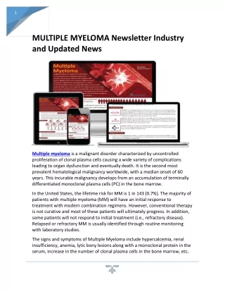
MM - Background • Multiple myeloma : as myeloma or plasma cell myeloma • cancer of the plasma cell • Multiple myeloma • excessive numbers of abnormal plasma cells in the bone marrow • overproduction of intact monoclonal immunoglobulin (IgG, IgA, IgD, or IgE) or Bence-Jones protein (free monoclonal κ and λ light chains)
MM - Background • Normal Plasma Cell Function in the Immune System • Stem cells can develop into B lymphocytes -- >travel to the lymph nodes, mature, and then travel throughout the body. • When foreign substances (antigens) enter the body -- >B cells develop into plasma cells that produce immunoglobulins Ig (antibodies) to help fight infection and disease.
Figure legend: In multiple myeloma, the B cell is damaged and gives rise to too many plasma cells (myeloma cells). These malignant cells do not function properly and their increased numbers produce excess immunoglobulins of a single type that the body does not need along with reduced amounts of normal immunoglobulins.
MM – Epidemiology (1) • Second most prevalent blood cancer • Approximately 1% of all cancers and 2% of all cancer deaths. • 45.000 currently have multiple myeloma • 14.600 new cases of myeloma each year. • Responsible for more than 10.000 deaths in the United States annually. • 5 year survival rate
MM – Epidemiology (2) MM occurs in all races and all geographic locations African Americans and blacks from Africa is two to three times the risk in whites Risk is lower in Asians from Japan and in Mexicans Slightly more frequent in men than in women (1.4:1)
MM – Epidemiology (3) MM is a disease of older adults The median age at diagnosis is 66 years Only 10 percent of patients are younger than 50 years Only 2 percent of patients are younger than 40 years
MM – Etiology • Genetic causes • Ongoing research is investigating whether HLA-Cw5 or HLA-Cw2 may play a role in the pathogenesis of myeloma. • Environmental or occupational causes • significant exposures in the agriculture, food, silicon, Benzene, Nikel and petrochemical industries • Radiation: • Radiation has been linked to the development of myeloma. • In 109,000 survivors of the bombing of Nagasaki, 29 died from myeloma from 1950-1976; however, some recent studies do not confirm that these survivors have an increased risk of developing myeloma.
MM - Pathophysiology • These myeloma cells travel through the bloodstream and collect in the bone marrow, where they cause permanent damage to healthy tissue. • As tumors grow, they invade the hard outer part of the bone, the solid tissue. • In most cases, the myeloma cells spread into the cavities of all the large bones of the body, forming multiple small lesions. This is why the disease is known as "multiple" myeloma.
Figure legend: Bone marrow stromal cells and myeloma cells produce cytokines that help myeloma cells grow and survive. Myeloma cells also produce growth factors that stimulate new blood vessel formation through a process called angiogenesis. New blood vessels provide nutrients and oxygen to the tumor, allowing it to grow. The natural immune response that attacks myeloma cells is suppressed.
Causes Bone pains Bone lesions Deformari Fractures on pathologic bone BONE MARROW MONOCLONAL PLASMA CELL PROLIFERATION Involvment of immune system Hypercalcemia Cytopenia Increased infectious risk Hypervascosity Hyperproduction of monoclonal component • Auto-Antibody activity • eritrocite - hemaglutininelor la rece • trombocite – afectarea functii • mielina - neuropatie senzitivo-motorie • factori ai coagulare - hemoragii • factorul von Willebrand • lipoproteina - hiperlipemii si xantoame • hormoni tiroidieni - hipotiroidie • structuri ale peretelui vascular Renal clearance Renal insuficiency
MM - Pathophysiology • Normally, plasma cells produce immunoglobulins to fight infection • However, in MM and MGUS a single cloned plasma cell proliferate and overproduce the same Ig (aka, the "M-protein" or "paraprotein." ) • The M-protein is usually an IgG • MM cells can also just produce the light chain component (Instead of the entire Ig)
MM - Pathophysiology • 80% of cases of MM arise De Novo • 20% percent from MGUS. • Risk factors for progression from MGUS to MM include: • An elevated M protein level > 1.5 g per dL • A non-IgG MGUS • Abnormal free light chain ratio • Patients with MGUS should be monitored closely q 6 to 12 months. (C-Level)
MM: Clinical Presentations Anemia - 73 percent Bone pain - 58 percent Elevated creatinine - 48 percent Fatigue/generalized weakness - 32 percent Hypercalcemia- 28 percent Weight loss - 24 percent, one-half of whom had lost ≥ 9 kg
MM – Clinical presentation Clinical manifestations are related to malignant behavior of plasma cells and abnormalities produced by M protein • plasma cell proliferation: multiple osteolytic bone lesions hypercalcemia bone marrow suppression ( pancytopenia ) • monoclonal M protein decreased level of normal immunoglobulins hyperviscosity
MM – Clinical presentation • Bone pain • Myeloma bone disease proliferation of tumor cells and release of IL-6 (osteoclast activating factor :OAF) stimulates osteoclasts to break down bone leading to hypercalcemia • These bone lesions in plain radiographs-- > "punched-out"/ lytic bone lesion
MM – Clinical presentation • Bone pain • Myeloma bone pain involves the rib ,sternum, spine , clavicle , skull , humerus & femur • The lumbar vertebrae are one of the most common sites of pain -- >may lead to spinal cord compression. • Persistent localized pain may indicate a pathological fracture.
MM – Clinical presentation • Hypercalcemia - patients present with confusion, somnolence, bone pain, constipation, nausea, and thirst. • Medical emergency • Anemia - normocytic and normochromic. • It results from the replacement of normal bone marrow by infiltrating tumor cells and inhibition of normal red blood cell production by cytokines.
MM – Clinical presentation • Bleeding • bleeding resulting from thrombocytopenia. • In some patients, monoclonal protein may absorb clotting factors and lead to bleeding, but this development is rare. • Hyperviscosity • high volume of monoclonal protein blood viscosity increases complications such as stroke, myocardial ischemia, or infarction. • Depends on the physical properties of the M component (most common with IgM, IgG, and IgA paraproteins). • Paraprotein concentrations of ~40 g/L (4 g/dL) for IgM, 50 g/L (5 g/dL) for IgG3, and 70 g/L (7 g/dL) for IgA. • Symptoms: headache, fatigue, visual disturbances, and retinopathy • Medical emergency
MM – Clinical presentation • Infection • Organism : polysaccharide encapsulated <strep.pneumoniae, H.influenzae> • Common pneumonia pathogens :S pneumoniae, S aureus, and K pneumoniae • Common pathogens causing pyelonephritis : E coli and other gram-negative organisms. • The increased risk of infection is due to immune deficiency resulting from diffuse hypogammaglobulinemia, which is due to decreased production and increased destruction of normal antibodies.
MM – Clinical presentation Renal failure - Occurs in nearly 25% of myeloma patients, • Hypercalcemia • Glomerular deposits of amyloid, • hyperuricemia, • recurrent infections, f • requent use of nonsteroidal anti-inflammatory agents for pain control, use of iodinated contrast dye for imaging, bisphosphonate use, myeloma cells infiltrates • Tubular damage associated with the excretion of light chains
MM – Laboratory tests • ESR > 100 • anaemia, thrombocytopenia • rouleaux in peripheral blood smears • marrow plasmacytosis > 10 -15% • hyperproteinemia • hypercalcemia • proteinuria • azotemia • osteolytic lesions in bones
MM – Laboratory tests • total protein, albumin and globulin, BUN, creatinine, and uric acid, which is high if the patient has high cell turnover or is dehydrated • Serum protein electrophoresis, urine protein electrophoresis, and immunofixation • Serum protein electrophoresis is used to determine the type of each protein present and may indicate a characteristic curve (ie, where the spike is observed). • Urine protein electrophoresis is used to identify the presence of the Bence Jones protein in urine. • Immunofixation is used to identify the subtype of protein (ie, IgA lambda).
Immunofixation to Determine Type of Monoclonal Protein IgG kappa M protein Lambda Light Chains Kyle RA and Rajkumar SV. Cecil Textbook of Medicine, 22nd Edition, 2004
MM – Laboratory tests • A 24-hour urine collection for the Bence Jones protein (ie, lambda light chains), protein, and creatinine • Quantification of proteinuria is useful for diagnosis (>1 g of protein in 24 h is a major criterion) and for monitoring the patient's response to therapy. • Creatinine clearance can be useful for defining the severity of the patient's renal impairment.
MM - Imaging Work up • Skeletal Survey • Skull, spine, long bones, ribs, pelvis • Diffuse osteopenia may suggest myelomatous involvement before discrete lytic lesions are apparent. • Do not use bone scans to evaluate myeloma • MRI • More sensitive • But, generally reserved for suspected spinal lesions
MM – Laboratory tests • Procedures • bone marrow aspirate & biopsy • samples to calculate the percent of plasma cells in the aspirate (reference range, <3%) and to look for sheets or clusters of plasma cells in the biopsy specimen.
Bone marrow aspirate : plasma cells of multiple myeloma.Note the blue cytoplasm, eccentric nucleus, and perinuclear pale zone (or halo).
M M - Diagnostic Criteria Major criteria I. Plasmacytoma on tissue biopsy II. Bone marrow plasma cell > 30% III. Monoclonal M spike on electrophoresis IgG > 3,5g/dl, IgA > 2g/dl, light chain > 1g/dl in 24h urine sample Minor criteria a. Bone marrow plasma cells 10-30% b. M spike but less than above c. Lytic bone lesions d. Normal IgM < 50mg, IgA < 100mg, IgG < 600mg/dl
M M - Diagnostic Criteria Diagnosis: • I + b, I + c, I + d • II + b, II + c, II + d • III + a, III + c, I II + d • a + b + c, a +b + d
MM - Staging Clinical staging • is based on level of haemoglobin, serum calcium, immunoglobulins and presence or not of lytic bone lesions • correlates with myeloma burden and prognosis I. Low tumor mass II. Intermediate tumor mass III. High tumor mass • subclassification A - creatinine < 2mg/dl B - creatinine > 2mg/dl
MM - Staging • Durie-Salmon staging system • High tumor mass <stage III > one of following abnormalitie mus be present • Hb <8.5 g/dl, Hct < 25 % • Sr Ca > 12 gm/dl • Very high Sr or Urine myeloma protein production rate 1. Ig G peak >7 gm/dl 2. IgA peak > 5 gm/dl 3. Bence Joneprotein > 12 gm/ 24 hr • > 3 lytic bone lesion on bone survey
MM - Staging • Durie-Salmon staging system 2. Low tumor mass <stage I> all of following must be present • Hb > 15 gm/dl, Hct> 32% • Sr Ca normal • Low Sr myeloma protein production rate • 1. Ig G peak< 5 gm/dl • 2. IgA peak < 3 gm/dl • 3. Bence Jone protien < 4 g/ 24 hr • No bone lesion or osteoporosis
MM - Staging • Durie-Salmon staging system 3.Intermediate tumor mass <stage II> a. no renal failure <Cr < 2 mg/dl> b. Renal failure <Cr > 2 mg/dl>
MM - Staging • The International Staging System (ISS) • Stage I : β2-microglobulin (β2M) < 3.5 mg/L, albumin > 3.5 g/dL • Stage II : β2M < 3.5 and albumin < 3.5; or β2M between 3.5 and 5.5 • Stage III : β2M > 5.5 Median overall survival for patients with ISS stages I, II, and III are 62, 44, and 29 months
MM: Treatment Decisions Indications for treatment Risk stratification Eligibility for stem cell transplantation
MM: Treatment • Active care • Chemotherapy • Autologous / Allogenic stem cell transplamtation • Drug : Arsenic trioxide, Thalidomide & Immunomodulator • Interferon • Supportive care • Radiation therapy • Bisphosphonate • Kayphoplasty
Smoldering (asymptomatic) myeloma Deferral of chemotherapy until progression to symptomatic disease Follow these patients closely, every 3 to 4 months, with serum protein electrophoresis, complete blood count, serum creatinine, and serum calcium Metastatic bone survey should be considered annually because asymptomatic bone lesions may develop
MM: Indications for Treatment Anemia (hemoglobin <10 g/dL or 2 g/dL below normal) Hypercalcemia (serum calcium >11.5 mg/dL) Renal insufficiency (serum creatinine>2 mg/dL) Lytic bone lesions or severe osteopenia Extramedullary plasmacytoma
MM: Risk stratification FISH for detection of t(4;14), t(14;16), and del17p13 Conventional cytogenetics (karyotyping) for detection of del 13 or hypodiploidy The presence of any of the above markers defines high risk myeloma, which encompasses the 25 percent of MM patients who have a median survival of approximately two years or less despite standard treatment
Current Frontline Options Conventional chemotherapy Survival ≤ 3 yrs Transplantation Prolongs survival 4-5 yrs Novel agents targeting stromal interactions and associated signaling pathways have shown promise Chng WJ, et al. Cancer Control. 2005;12:91-104.
MM: initial therapy The initial therapy of patients with symptomatic myeloma varies depending on whether patients are eligible or not to pursue autologous hematopoietic cell transplantation