Download
1 / 48
580 likes | 1.24k Views
Diagnosis and Management of Pelvic Organ Prolapse. Leslie Ablard M.D. Pelvic Organ Prolapse (POP) Herniation of the pelvic organs to or beyond the vaginal walls Annual cost of ambulatory care from 2005 to 2006 was almost $300 million

E N D
Diagnosis and Management of Pelvic Organ Prolapse Leslie Ablard M.D.
Pelvic Organ Prolapse (POP) • Herniation of the pelvic organs to or beyond the vaginal walls • Annual cost of ambulatory care from 2005 to 2006 was almost $300 million • Surgical repair of prolapse was the most common inpatient procedure performed in women older than 70 yrs from 1979 to 2006 • Approximately 11% of all women will undergo surgical repair for POP or incontinence by age 80
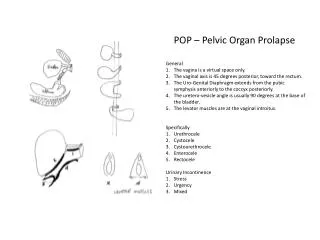
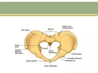
Terminology • Anterior compartment prolapse (cystocele) • Hernia of anterior vaginal wall often associated with descent of the bladder • Posterior compartment prolapse (Rectocele) • Hernia of the posterior vaginal segment often associated with descent of the rectum • Apical compartment prolapse (uterine prolapse, vaginal vault prolapse) • Descent of the apex of the vagina into the lower vagina, to the hymen, or beyond the vaginal introitus • The apex can be either the uterus and cervix, cervix alone, or vaginal vault • Apical prolapse is often associated with enterocele. • Enterocele • Hernia of the intestines to or through the vaginal wall
Terminology • Procidentia • Hernia of all three compartments through the vaginal introitus.
Terminology • The terms anterior vaginal wall prolapse and posterior vaginal wall prolapse are preferred to cystocele and rectocele because vaginal topography does not reliably predict the location of the associated viscera in POP • Division of the vagina into separate compartments is somewhat arbitrary, because the vagina is a continuous organ and prolapse of one compartment is often associated with prolapse of another • As an example, approximately half of anterior prolapse can be attributed to apical descent
Risk Factors • Parity — The risk of POP increases with increasing parity • Prospective cohort study of more than 17,000 • The risk of hospital admission for POP increased • 1st birth- 4-fold • 2nd - 8-fold • 3rd - 9-fold • 4th- 10-fold • Among parous women, it has been estimated that 75 percent of prolapse can be attributed to pregnancy and childbirth • Advancing Age- Older women are at increased risk for POP • Every additional 10 yrs of age increased prolapse risk by 40%
Risk Factors • Obesity • Overweight and obese women (body mass index >25) have a two-fold higher risk of having prolapse than other women • Hysterectomy • Hysterectomy is associated with increased apical prolapse • ? Vaginal > Abdominal ? • Other risk factors • Chronic constipation is a risk factor for POP, likely due to repetitive increases in intraabdominal pressure • COPD, etc conditions that also increase intraabdominal pressure
Risk Factors • Race and Ethnicity- • African Americans lower prevalence than other ethnic groups • Risk of Latina and white women is four to five fold higher than AA
Clinical Manifestations • Patients may present with symptoms related specifically to the prolapsed structures • bulge or vaginal pressure or with associated symptoms including urinary, defecatory or sexual dysfunction • Symptoms such as low back or pelvic pain have often been attributed to POP, but this association is not supported by well-designed studies • Severity of symptoms does not correlate well with the stage of prolapse
Clinical Manifestations • Symptoms are often related to position; they are often less noticeable in the morning or while supine and worsen as the day progresses. • Many women with prolapse are asymptomatic; treatment is generally not indicated in these women.
Clinical Manifestations • Bulge Symptoms • In a study of 1912 women presenting to a pelvic floor disorder clinic, symptoms of “a bulge or that something is falling out of the vagina” had a sensitivity of 67 percent and a specificity of 87 percent for POP at or past the hymen • Although complaints of a bulge are associated with the presence of prolapse, it is only weakly correlated with prolapse stage, and does not predict site of prolapse • Protrusion from the vagina may cause chronic discharge and/or bleeding from ulceration
Urinary Symptoms • Loss of support of the anterior vaginal wall or vaginal apex may affect bladder and/or urethral function. • Symptoms of stress urinary incontinence (SUI) often coexist with stage I or II prolapse • As prolapse advances, women may experience improvement in SUI, but increased difficulty voiding • Advanced anterior or apical prolapse may “kink” the urethra and result in symptoms of obstructed voiding such as • slow urine stream • need to change position • manually reduce (splint) the prolapse to urinate • sensation of incomplete emptying • complete urinary retention
Urinary Symptoms • 13% to 65% of continent women develop symptoms of SUI after surgical correction of prolapse • Elevation of prolapse during pelvic examination with prolapse treatment may unmask “occult” SUI • Women with POP have a two- to five-fold risk of overactive bladder symptoms (urgency, urge urinary incontinence, frequency) compared with the general population
Diagnosis and Classification • To POP-Q or not to POP-Q • POPQ system The POPQ system is an objective, site-specific system for describing and staging POP in women • The POPQ system involves quantitative measurements of various points representing anterior, apical, and posterior vaginal prolapse to create a "topographic" map of the vagina • These anatomic points can then be used to determine the stage of the prolapse
Staging • Stage 0- No prolapse • Aa, Ba, Ap, Bp are -3 cm and C or D≤ -(tvl - 2) cm • Stage 1- Most distal portion of the prolapse -1 cm (above the level of hymen) • Stage 2- Most distal portion of the prolapse ≥ -1 cm but ≤ +1 cm (≤1 cm above or below the hymen) • Stage 3 - Most distal portion of the prolapse > +1 cm but < +(tvl - 2) cm (beyond the hymen; protrudes no farther than 2 cm less than the total vaginal length) • Stage 4 - Complete eversion; most distal portion of the prolapse ≥ + (tvl - 2) cm
Why POP-Q • The POPQ has proven interobserver and intraobserver reliability • The POPQ system is the POP classification system of choice of the International Continence Society (ICS), the American Urogynecologic Society (AUGS), and the Society of Gynecologic Surgeons • It is the system used most commonly in the medical literature
Baden-Walker System • The Baden-Walker Halfway Scoring System is the next most commonly used POP staging system • The degree, or grade, of each prolapsed structure is described individually • The grade/degree is defined as the extent of prolapse for each structure noted on examination while the patient is straining • The Baden-Walker system lacks the precision and reproducibility of the POPQ system
Baden-Walker System • The system has five degrees/grades • 0 – No prolapse • 1 – Leading edge of prolapsed structure descends halfway to vaginal introitus (hymen) • 2 – Leading edge of prolapsed structure descends to the vaginal introitus • 3 – Leading edge of prolapsed structure(s) protrudes up to halfway outside the vagina • 4 – Leading edge of prolapsed structure(s) protrudes more than halfway outside the vagina
Examination • Examination components • Visual inspection • Speculum examination • Bimanual pelvic examination • Rectovaginal examination • Pelvic Floor Muscle evaluation
Equipment • Instruments • Sims retractor (single blade speculum) or a bivalve speculum that can be easily taken apart so that the anterior and posterior blades can be used separately to observe individual compartments of the vagina (anterior, posterior, apical). • To make the measurements for the POPQ system, a ruler or a large cotton swab or sponge forceps marked in 1 cm increments is used • Ring Forceps occasionally used for evaluation of occult incontinence to reduce prolapse
Patient Positioning ??? • The examination is performed with resting and maximal straining position • The patient is examined initially in the dorsal lithotomy position • The examination is then repeated with the patient standing • In the standing position, the patient places one foot on a well-supported footstool. The examining gown is lifted slightly to expose the genital area during the examination
Visual Inspection • The first part of the examination is a visual inspection of the vulvar, perineal, and perianal areas with the patient in the dorsal lithotomy position • As during other components of the examination, the inspection should be performed initially with the patient relaxed and then while straining • Findings that should be noted during this component of the examination include: • Transverse diameter of the genital hiatus (eg, the space between the labia majora) • Protrusion of the vaginal walls or cervix to or beyond the introitus (procidentia) • Length and condition of the perineum • Rectal prolapse • In patients with prolapse to or beyond the hymen, the vaginal tissue is examined for ulceration. • Any other findings (eg, skin or mucosal lesions) should be noted and evaluated appropriately
Speculum and Bimanual Exam • The speculum and bimanual examinations are the principal components • Prolapse of each anatomic compartment is evaluated as follows: • Apical prolapse (prolapse of the cervix or vaginal vault) – A bivalve speculum is inserted into the vagina and then slowly withdrawn; any descent of the apex is noted • Anterior vaginal wall – A Sims retractor or the posterior blade of a bivalve speculum is inserted into the vagina with gentle pressure on the posterior vaginal wall to isolate visualization of the anterior vaginal wall • Posterior vaginal wall – A Sims retractor or the posterior blade of a bivalve speculum into the vagina with gentle pressure on the anterior vaginal wall to isolate visualization of the posterior vaginal wall • To complete the exam, a bimanual examination is performed in order to evaluate for any coexisting pelvic abnormalities
Rectovaginal Examination • Diagnose an enterocele • Differentiate between a high rectocele and an enterocele • Assess the integrity of the perineal body • Detect rectal prolapse • The best method for detecting an enterocele is to perform the rectovaginal exam with the patient standing (?); the small bowel can be palpated in the cul-de-sac between thumb and forefinger
Neurologic/Pelvic Floor Muscle Evaluation • Pelvic floor muscle testing • The pelvic floor musculature is inspected to evaluate integrity and symmetry • The examiner should also note the presence of scarring and whether pelvic floor contraction pulls the perineum inward • Palpation through the vagina or rectum helps in assessing pelvic floor squeeze strength and levator muscle thickness. • The tone and strength of the pelvic floor muscles can be assessed by asking the patient to contract the pelvic floor muscles around the examining fingers. • Women with poor pelvic floor muscle function may benefit from pelvic physical therapy
Treatment • Establishing patient goals • Treatment is individualized according to each patient’s symptoms and their impact on her quality of life • Patient satisfaction after POP surgery correlates highly with achievement of self-described, preoperative surgical goals, but poorly with objective outcome measures • Management options • Women with symptomatic prolapse can be managed expectantly, or treated with conservative or surgical therapy • Both conservative and surgical treatment options should be offered. • There are no high quality data comparing these two approaches
Treatment • Physical Therapy- • Pelvic floor muscle exercises (PFME) appears to improve stage and symptoms • The best designed randomized trial included 109 women with stage I to III prolapse who were assigned to either PFME for six months or control group • Women in the PFME group had significant reductions in the frequency and bother of most prolapse, bladder, and bowel symptoms (exceptions were urge urinary incontinence symptoms, difficulty with stool emptying, and solid stool fecal incontinence) • Improvement in POP stage was found more frequently in the PFME group (19 versus 8 percent)
Treatment • Estrogen therapy ? • Use of estrogen and estrogenic agents (raloxifene) appears to be associated with a decrease in undergoing surgery for POP, according to a systematic review of randomized trials • This systematic review included six trials, however, none of these evaluated the role of estrogen in treating POP
Treatment • Vaginal pessary • The mainstay of non-surgical treatment for POP is the vaginal pessary • Pessaries are silicone devices in a variety of shapes and sizes, which support the pelvic organs • Approximately half of the women who use a pessary continue to do so in the intermediate term of one to two years • Pessaries must be removed and cleaned on a regular basis • CONTRAINDICATIONS • Local infection — Active infections of the vagina or pelvis, such as vaginitis or pelvic inflammatory disease, preclude the use of a pessary until the infection has been resolved • Latex sensitivity — The Inflatoball pessary is made of latex; therefore, it is contraindicated in women with latex allergies. The other pessaries discussed below are nonallergenic. • Noncompliance — Noncompliance with follow-up could be harmful since an undetected and untreated erosion could put the patient at risk of developing a fistula • Sexually active women who are unable to remove and reinsert the pessary — Inability to manage the pessary around coital activity could be discouraging
Treatment • Fitting the pessary • Women to be fitted for a pessary are first examined with an empty bladder in the dorsal lithotomy position • Pessaries are inserted into the vagina with the dominant hand, while the nondominant hand separates the introitus and depresses the perineal body. • After the pessary is inserted into the vagina, the woman is asked to strain and cough repeatedly on the examination table, ambulate in the office, and void and strain while sitting on a toilet • This "office trial" helps determine if she will be able to retain the pessary and void when she returns home, and if bothersome urinary incontinence will develop. • She should have a negative cough stress test following pessary placement, as she is unlikely to be satisfied if there are significant SUI symptoms • Women should be reassured that it is not an emergency if the pessary is expelled; they should just bring the pessary back to the office and a different type or size of pessary will likely be effective
Follow-up • A follow-up visit is scheduled one to two weeks later. • The pessary is removed and cleaned with soap and water, and the vagina is examined for erosions • If the pessary fits well and there were no side effects, motivated and able patients are taught how to remove, clean, and reinsert their pessary at least once per week, with follow-up in one to two months, and every 6 to 12 months thereafter • If the patient cannot, or chooses not, to remove and reinsert her pessary, then she returns for follow-up in one to two months, and every three to four months thereafter for pessary cleaning and assessment by the provider.
Treatment • Offer most women low-dose estrogen vaginal cream (0.25 to 0.5 g applicator, two to three nights per week) to treat co-existing vaginal atrophy and dryness from estrogen deficiency • KY or other non-hormonal lubrication may be used for those patients where estrogen is contraindicated (breast ca, etc) • In some women, the width of the introitus may decrease in size after several weeks of pessary use. In such women, a new smaller size pessary is prescribed to allow for easier removal and insertion
Treatment-Surgical • Candidates • Symptomatic POP • Failed or declined conservative management • Women finished with childbearing • Reports of uterine sparing procedures • Young or Elderly- • Risk of recurrence in young (sacral colpopexy) and comorbidities in elderly (colpocliesis)
Treatment- Surgical • Reconstructive or obliterative • Most women with symptomatic POP are treated with a reconstructive procedure • Obliterative procedures (eg, colpocleisis) are reserved for women who cannot tolerate more extensive surgery or who are not planning future vaginal intercourse • Concomitant hysterectomy • When apical prolapse is repaired, the decision must be made whether to perform a hysterectomy as a part of the procedure.
Treatment- Surgical • Surgical route for repair of multiple sites of prolapse • Reconstructive surgery for POP often involves repair of multiple anatomic sites of prolapse (apical, anterior, posterior) • The choice of surgical route depends upon the optimal approach for the combination of prolapse sites. • Concomitant anti-incontinence surgery • Symptomatic POP often coexists with SUI and, in some women, anal incontinence • POP repair must be coordinated with treatment of incontinence. • Use of surgical mesh • Surgical mesh is used in abdominal POP repair • Use in transvaginal procedures has increased, but questions have arisen about the safety of this approach.


![get [PDF] Download Pelvic Organ Prolapse: The Silent Epidemic download](https://cdn7.slideserve.com/12583059/pelvic-organ-prolapse-the-silent-epidemic-dt.jpg)


