Understanding the Kidney's Role in Homeostasis and Hormone Production
The kidney plays a critical role in maintaining water, salt, and electrolyte balance while functioning as an endocrine gland. It secretes hormones vital for regulating blood pressure and producing red blood cells. The kidneys filter approximately 1 liter of blood per minute, adjusting blood composition and eliminating metabolic waste. They perform glomerular filtration and renal reabsorption, affecting various substances like glucose and electrolytes. The activation of vitamin D and the release of erythropoietin showcase the kidney's importance in maintaining plasma calcium levels and red blood cell production.

Understanding the Kidney's Role in Homeostasis and Hormone Production
E N D
Presentation Transcript
Chapter 16 The Genitourinary System The kidney is essential in maintaining water, salt, and electrolyte balance and is an endocrine gland that secretes at least three hormones. The kidney helps control blood pressure and is especially susceptible to damage if blood pressure is too high or too low. Physiologic Concepts Renal Blood Flow The kidneys receive approximately 1 L of blood per minute,one-fifth of the cardiac output. This high rate of blood flow is for allowing the kidney to adjust the blood composition continually. By adjusting the blood composition, the kidney is able to maintain blood volume; ensure sodium, chloride, potassium, calcium, phosphate, and pH balance; and eliminate products of metabolism such as urea and creatinine.
Glomerular Filtration • The process of filtration across the glomerulus is similar to that which occurs across all capillaries but the glomerular capillaries have increased permeability to small solutes and water. Also, unlike other capillaries, the forces favoring filtration of plasma across the glomerular capillary into Bowman's space are greater than the forces favoring reabsorption of fluid back into the capillary. Therefore, net filtration of fluid into Bowman's space occurs. This fluid then diffuses into Bowman's capsule and begins its journey through the rest of the nephron.
Renal Reabsorption • Reabsorption is the second process by which the kidney determines the concentration of a substance filtered from the plasma. Reabsorption refers to the active (requiring energy and always being mediated by a carrier) or the passive (no energy required) movement of a substance filtered at the glomerulus back into the peritubular capillaries. Reabsorption may be total (e.g., glucose) or partial (e.g., sodium, urea, chloride, and water). Renal Endocrine Function 1-The kidney functions as an endocrine organ, not only with the production and release of renin but also with the production and release of two other hormones: 1,25-dihydroxyvitamin D3, important for bone mineralization; and erythropoietin, required for red blood cell production.
2- 1,25-Dihydroxyvitamin D3 The kidney acts in conjunction with the liver to produce an active form of vitamin D, called 1,25-dihydroxyvitamin D3, from an inactive precursor consumed in the diet. The inactive form of vitamin D can also be produced in a reaction catalyzed by sunlight on a precursor present in the skin. Vitamin D is essential for maintenance of plasma calcium levels required for bone formation. The active form of vitamin D acts as a hormone by circulating in the blood and stimulating absorption of calcium and, to a lesser extent, phosphate across the small intestine and across the kidney tubules. Vitamin D also stimulates bone resorption (breakdown). Bone resorption releases calcium, and thus plasma calcium is increased by this mechanism as well.
Parathyroid hormone is the stimulus for the kidney to play its role in activating vitamin D3. Parathyroid hormone is released from the parathyroid gland in response to decreased plasma calcium. This is an example of a negative feedback cycle: decreased plasma calcium leads to increased parathyroid hormone, which leads to increased renal activation of vitamin D3. • Activation of vitamin D3 increases gut and kidney absorption of calcium, increasing plasma calcium and removing the stimulus for parathyroid release. Parathyroid hormone also directly stimulates bone resorption to release calcium into the plasma when necessary. Individuals who have renal disease frequently develop brittle, easily broken bones as a result of too little active vitamin D3.
3- Erythropoietin • The hormone that stimulates the bone marrow to increase the production of erythrocytes (red blood cells) is called erythropoietin. The cells of the kidney responsible for synthesizing and releasing erythropoietin respond to renal hypoxia. Individuals who have renal disease frequently demonstrate chronic and debilitating anemia.
Micturition • Micturition is the process of urination, which is the elimination of urine from the body. Micturition occurs when the internal and the external urethral sphincters at the base of the bladder are relaxed. The bladder is composed of smooth muscle (the detrusor muscle), innervated by sensory neurons that respond to stretch, and parasympathetic fibers that travel from the sacral area to the bladder. An area of smooth muscle at the base of the bladder (the internal sphincter) is also innervated by parasympathetic nerves. An external sphincter composed of skeletal muscle is just below the internal sphincter and at the top of the urethra. When urine accumulates, stretch of the bladder is sensed by afferent fibers that send the information to the spinal cord. Parasympathetic nerves to the bladder are activated, causing contraction of the smooth muscle and opening of the internal sphincter. At the same time, the motor neurons going to the external sphincter are inhibited and the external sphincter is relaxed, causing micturition to occur.
Micturition, however, can be voluntarily inhibited. This is possible because at the same time that the afferent nerves are conveying information on bladder stretch to the spinal cord, they are also sending information up the cord to the brainstem and cortex, allowing one to be conscious of the need to void. Descending neurons from the brain can inhibit or stimulate the spinal reflex to void. These descending pathways inhibit urination by causing contraction of the skeletal muscles of the pelvis as well as the external sphincter. Descending pathways also block the firing of parasympathetic nerves to the internal sphincter. For urination to be facilitated, skeletal muscles can be voluntarily relaxed. Voluntary control over micturition becomes functional in children by or before the time they become 3 or 4 years of age. However, it may become interrupted at any time by central nervous system disease or injury or from spinal cord trauma.
Tests of Renal Function • Blood Urea Nitrogen Urea is a nitrogenous waste product of protein and amino acid metabolism. One important job of the kidney is to eliminate this potentially toxic substance from the body. With declining renal function, blood urea nitrogen (BUN) levels increase. Measuring BUN therefore provides an indication of kidney health. • BUN, however, is not only determined by renal function. It can also be affected by circumstances not associated with the kidney, such as increased or decreased dietary protein intake, or any unusual cause of an increased protein breakdown, such as a muscle injury. Likewise, liver disease may decrease BUN, because the liver is necessary to convert ammonia to urea
. Because BUN levels are affected by these other factors, BUN level alone may be an indiscriminate indicator of renal disease. Therefore, often the ratio of BUN to serum creatinine is reported as well. Normally BUN and creatinine co-vary, keeping this ratio at approximately 10:1. If BUN is affected by other than renal factors, however, this ratio may change. Ratios greater than 15:1 suggest a non-renal cause of urea elevation. Ratios less than 10:1 occur with liver disease
Serum Creatinine Creatinineis a product of muscle breakdown. Creatinine is excreted by the kidney through a combination of filtration and secretion. The concentration of creatinine in the plasma remains nearly constant from day to day. It varies slightly from approximately 0.7 mg/100 mL of blood in a small woman to 1.5 mg/l00 mL in a muscular man. Levels greater than these suggest the kidney is not clearing creatinine and indicate renal disease. Serum creatinine is very indicative of renal function. As a rough guide, a doubling of serum creatinine levels indicates a 50% reduction in renal function. Likewise, a tripling of normal creatinine levels indicates a 75% reduction in renal function.
Urinalysis A urine sample may be easily obtained and evaluated for the presence of red blood cells, protein, glucose, and leukocytes, all of which are normally minimal to absent in the urine. Urine casts, which occur in the presence of high amounts of urine protein, may also be observed under some conditions of renal disease or injury. Urine osmolality (specific gravity) is measurable and should range between 1.015 and 1.025. Dehydration causes increased urine osmolality as more water is reabsorbed back into the peritubular capillaries. Overhydration results in decreased urine osmolality.
Cystoscopy Cystoscopyis the process in which a lighted scope (cystoscope) is inserted up the urethra into the bladder. Bladder lesions, stones, and biopsy samples may be taken. Voiding Cystourethrography Voiding cystourethrography involves bladder catheterization and infusion of a radioactive dye to study the shape and size of the bladder. It can be used to detect and grade the degree of vesicoureteral reflux. If used inappropriately, cystourethrography may spread an unresolved bladder infection into the ureters or kidney.
Intravenous Urography Intravenous urography is a technique in which a radiologic dye is injected intravenously, and x-ray films are taken sequentially as the dye filters through the kidney. Obstructions to flow in the glomeruli or tubules, vesicoureteral reflux, and stones may be visualized. A drawback to the use of this technique is the finding that some individuals are allergic to the dye and may suffer an anaphylactic reaction. High doses of radiation are involved.
Renal Ultrasound Renal ultrasound uses the reflection of sound waves to identify renal abnormalities, including structural abnormalities, kidney stones, tumors, and other masses. Because it is non-invasive and does not involve radiation exposure, this technique is frequently used to evaluate renal function in children who have had a urinary tract infection. It does not, however, offer sufficient detail to evaluate vesicoureteral reflux, renal scarring, or inflammation.
Pathophysiologic Concepts Alterations in Glomerular Filtration Glomerular filtration depends upon the summation of forces favoring filtration of plasma out of the glomerulus and forces favoring reabsorption of filtrate into the glomerulus. Anything that affects the forces of filtration or the forces of reabsorption affects net glomerular filtration. Forces favoring filtration are capillary pressure and interstitial fluid colloid osmotic pressure. Forces favoring reabsorption are interstitial fluid pressure and plasma colloid osmotic pressure
Tubular Obstruction One cause of increased interstitial fluid pressure is tubular obstruction. Obstruction present in the nephron causes fluid to back up into Bowman's capsule and the interstitial space. Unrelieved tubular obstruction can collapse the nephrons and capillaries and can lead to irreversible damage, especially to the renal papillae, which are the final site for urine concentration. Causes of obstruction include renal calculi (stones) and scarring from repeated kidney infections.
Azotemia Azotemia refers to abnormal elevation of nitrogenous waste products in the blood such as urea, uric acid, and creatinine. Azotemia indicates a decrease in GFR, occurring either acutely or with chronic renal failure. Azotemia is an early sign of renal damage. Uremia Uremia is not a single event, but rather a syndrome (a constellation of symptoms) that develops in an individual who has end-stage renal disease. Because the kidney is pivotal in maintaining water, acid-base, and electrolyte balance and in removing toxic waste products, the symptoms of uremia are widespread and affect all the organs and tissues of the body. Common symptoms include fatigue, anorexia, nausea, vomiting, and lethargy. Intractable itching (pruritus) may occur. Hypertension, osteodystrophy, and uremic encephalopathy develop as well, with central nervous system changes, including confusion and psychosis, characterizing end stages.
Nephrotic Syndrome Nephrotic syndrome is the loss of 3.5 g or more of protein in the urine per day. Under normal circumstances, virtually no protein is lost in the urine. Nephrotic syndrome usually indicates severe glomerular damage. Diabetic nephropathy is the most common cause of nephrotic syndrome. Clinical manifestations may include increased susceptibility to infections (caused by hypoimmunoglobulins) and generalized edema, called anasarca.
Anasarca Defined as a generalized edema in individuals suffering from hypoalbuminemia as a result of nephrotic syndrome or other conditions, anasarca is caused by a systemic decrease in capillary osmotic pressure. With a decrease in this major force favoring reabsorption of interstitial fluid back into the capillaries, edema of the interstitial space throughout the body occurs. The edema is usually soft and pitting and occurs early in the periorbital (surrounding the eye) regions, the ankles, and the feet.
Renal Osteodystrophy Demineralization of bone occurring with renal disease is known as renal osteodystrophy. Renal osteodystrophy has many causes, including decreased renal activation of vitamin D3, leading to decreased calcium absorption across the gut, and subsequent reduced serum calcium levels. Decreased serum calcium levels also stimulate parathyroid hormone release. An elevated bone breakdown contributes to easy bone fracturing.
Metabolic Acidosis/Renal Acidosis • Metabolic acidosis is a decrease in plasma pH not caused by a respiratory disorder. Chronic renal disease results in metabolic acidosis as a result of reduced H+ excretion and altered bicarbonate reabsorption. The result is increased plasma H+ and lowered pH. • The respiratory system is stimulated by the increase in hydrogen. Tachypnea (increased respiratory rate) occurs in an attempt to blow off the excess hydrogen as carbon dioxide. The respiratory response to renal acidosis is called respiratory compensation.
Uremic Encephalopathy Uremic encephalopathy refers to neurologic changes seen in severe renal disease. Symptoms include fatigue, drowsiness, lethargy, seizures, muscle twitching, peripheral neuropathy (pain in the legs and feet), decreases in memory, and coma. Uremic encephalopathy appears to be caused by accumulation of toxins, alterations in potassium balance, and decreased pH.
Renal Dialysis The process of adjusting blood levels of water and electrolytes in a person who has poor or non-functioning kidneys is called renal dialysis. In this procedure, blood is directed past an artificial medium containing water and electrolytes in predetermined concentrations. The artificial medium is the dialyzing fluid. By simple diffusion across a selectively permeable membrane, water and electrolytes in the blood move down their individual concentration gradients into or out of the dialyzing solution.Thereare two types of dialysis:
Hemodialysis Dialysis is performed outside the body. Blood is passed from the body, through an arterial catheter, into a large machine. Two chambers separated by a semipermeable membrane are inside the machine. Blood is delivered to one chamber, dialyzing fluid is placed in the other, and diffusion is allowed to occur. It takes about 3 to 5 hours and is required approximately three times per week. Hemodialysiscontributes to problems of anemia because some red blood cells are destroyed in the process. Infection is also a risk.
Peritoneal Dialysis The individual's own peritoneal membrane is used as a natural, semipermeable barrier. Prepared solution (approximately 2 L) is delivered into the peritoneal cavity. The solution is allowed to remain in the peritoneal cavity for a predetermined amount of time (usually between 4 and 6 hours). During this time, water and electrolytes diffuse back and forth between the circulating blood. The person can usually continue activity while the exchange takes place.
Kidney Transplantation Kidney transplantation involves placement of a donor kidney into the abdominal cavity of an individual suffering from end-stage renal disease. Transplanted kidneys can come from living or dead donors. The more similar the antigenic properties of the donated kidney are to the patient, the more likely the transplantation will be successful. With appropriate follow-up, approximately 94% of kidneys transplanted from cadavers and 98% from living donors function well after surgery. Long-term graft survival (10 years) is similar for both (approximately 78% for grafts from living donors versus 76% for grafts from cadavers). Individuals receiving kidney donation must remain on a variety of immunosuppressant medications for life to prevent organ rejection. I
Conditions of Disease or Injury Hypospadias Hypospadias is a congenital defect in males , the opening of the urethra is on the ventral side. This condition may be slight or extreme. Some infants demonstrate the urethral meatus (opening) in the scrotal or perineal area. Ejaculatory dysfunction in the adult male may occur. Treatment Surgical correction may be necessary, preferably before the child is 1 or 2 years old. Circumcision should be avoided in the newborn so that the foreskin may later be used for repair.
Renal agenesis • Failure of the kidneys to develop during gestation ,may be unilateral or bilateral. Bilateral agenesis is incompatible with life. • Unilateral agenesis results in hypertrophy of the remaining kidney as it adapts to compensate functionally for the absent kidney. Clinical Manifestations With unilateral renal agenesis, no symptoms are apparent if the remaining kidney is healthy. The remaining kidney may compensate and grow almost twice as big as otherwise expected. If the remaining kidney functions poorly, however, various disease manifestations may be present.
Diagnostic Tools • Prenatal ultrasound can often detect renal agenesis. • After birth, computerized axial tomography (CAT) scan or renal ultrasound is used to diagnose the condition. Treatment • No treatment is required for unilateral agenesis if the remaining kidney is healthy. • If structural or functional defects are present in the remaining kidney, surgery may be required.
Renal Calculi Renal calculi refer to stones that occur anywhere in the urinary tract. Calculi are most commonly made up of calcium crystals. Renal calculi can be caused by either increased urine pH (e.g., calcium carbonate stones) or decreased urine pH (e.g., uric acid stones). Anything that obstructs urine flow, leading to urine stasis anywhere in the urinary tract, increases the likelihood of stone formation.
Clinical Manifestations • Pain is often colicky (rhythmic), especially if the stone is in the ureter or below. The pain may be intense. The location of pain depends on the site of the stone. • A stone in the kidney itself may be asymptomatic unless it causes obstruction or an infection develops. • Hematuria, caused by irritation and injury of the renal structures, is common with calculi. • Decreased urine output results if obstruction to flow occurs.
Diagnostic Tools Radiograph, ultrasound, or intravenous urography may locate a stone. Complications • Urinary obstruction can lead to hydroureter, that is, abnormal distension of ureter with urine. Unrelieved hydroureter can lead to hydronephritis, swelling of the renal pelvis and collecting-duct system. leading to electrolyte and fluid imbalance. • Obstruction causes increased interstitial hydrostatic pressure and can lead to a decrease in GFR. Renal failure may develop if both kidneys are involved. • The chance of a bacterial infection increases. • Renal cancer may develop from repeated inflammation and injury.
Treatment • High fluid intake in individuals prone to calculi may prevent their formation. • Increased fluid intake increases urine flow and helps wash out the stone. • Appropriate alteration of urine pH may encourage stone breakdown. • Lithotripsy (shock wave therapy) or laser therapy may be used to break apart the stone. • Surgery may be necessary to remove a large stone or to place a diversion tube around the stone to relieve obstruction.
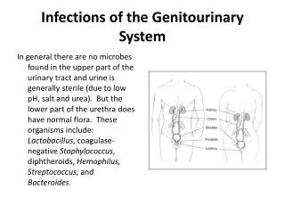
Urinary Tract Infection A urinary tract infection is an infection anywhere in the urinary tract, including the kidney itself. Most urinary tract infections are bacterial in origin, but fungi and viruses also may be implicated. The most common bacterial infection is by Escherichia coli, a fecal contaminant commonly found in the anal area. • Urinary tract infections are especially common in girls and women. One cause is the shorter urethra in the female, which allows the contaminating bacteria to gain access more easily to the bladder.
Individuals who have diabetes also are at risk of frequent urinary tract infections because of the high glucose content of the urine and poor immune function. • Persons who have a spinal cord injury or anyone using a urinary catheter to void are at increased risk of infection. Types of Urinary Tract Infections • Urinary tract infections may be divided into cystitis and pyelonephritis. Cystitis is an infection of the bladder, the most common site for an infection. Pyelonephritis is an infection of the kidney itself and can be either acute or chronic.
Acute pyelonephritis usually occurs as a result of an ascending bladder infection. It may also occur as a result of a blood-borne infection. Infections may be in both or in one kidney. • Chronic pyelonephritis may result from repeated infections and is usually found in individuals who have frequent calculi, other obstructions, or vesicoureteralreflux.With chronic pyelonephritis, extensive scarring and obstruction of the tubules result. The ability of the kidneys to concentrate urine decreases as tubules are lost. The glomeruli are usually unaffected. Chronic renal failure may develop.
Clinical Manifestations Cystitis typically presents with dysuria (pain on urination), increased frequency of urination, and a sense of urgency to urinate. • Lower back or suprapubic pain may occur, especially with pyleonephritis. • Fever accompanied by blood in the urine in severe cases. • Symptoms of infection in infants or young children may be non-specific and include irritability, fever, lack of appetite, vomiting, and very strong-smelling diapers.
Acute pyelonephritistypically presents with • Fever. • Chills. • Flank pain. • Dysuria. Chronic pyelonephritismay have manifestations similar to acute pyelonephritis. However, it can also include hypertension and may eventually lead to signs of renal failure.
Diagnostic Tools • Urine culture and sensitivity of the microorganism allow for identification and treatment. • White blood cells will be present in the urine with infection anywhere. White cell casts present in the urine suggest pyleonephritis rather than cystitis, since they indicate that white cells have been lysed in the tubules. Complications • Renal or perirenal abscess formation may occur. • Renal failure may develop after repeated infections if both kidneys are involved.
Treatment • Women and girls in particular should be encouraged to drink fluids frequently. • Girls should be taught at a young age to wipe from front to back after urination to avoid contamination of the urethral opening with fecal bacteria. • Women should be encouraged to urinate after sexual intercourse to wash out ascending microorganisms. • Antibiotic therapy with to repeat urinalysis during or after drug therapy is required. • If chronic pyelonephritis is caused by an obstruction or reflux, surgical treatment specific to relieve these problems is necessary.
Glomerulonephritis Glomerulonephritis is an inflammation of the glomerulus. Types of glomerulonephritis include : I-Acute Glomerulonephritis occurs as a result of deposition of antibody-antigen complexes in the glomerular capillaries. Complexes usually develop 7 to 10 days after a pharyngeal or skin streptococcal infection (poststreptococcalglomerulonephritis) but may follow any infection. An inflammatory reaction is initiated in the glomerulus after deposition of antibody-antigen complexes.
It usually resolves with specific antibiotic therapy, especially in children. II-Rapidly Progressive Glomerulonephritis Is an inflammation of the glomeruli that occurs so rapidly that there is a 50% decrease in GFR within 3 months of disease onset. Rapidly progressive glomerulonephritis can occur from a worsening of acute glomerulonephritis, from an autoimmune disease, or may be idiopathic (unknown) in origin.
III-Chronic Glomerulonephritis Is the long-term inflammation of the glomerular cells. It may occur as a result of unresolved acute glomerulonephritis, or it might develop spontaneously. It commonly occurs after years of subclinical glomerular injury and inflammation, associated with only slight hematuria and proteinuria. Clinical Manifestations All types of glomerulonephritis are associated with: • Decreased urine volume. • Blood in the urine (brownish-colored urine). • Fluid retention.
Diagnostic Tools • Hematuria as measured by urinalysis. • Red blood cell casts in the urine. • Proteinuriagreater than 3 to 5 g/day. • Decreased GFR as measured by creatinine clearance. • In poststreptococcalglomerulonephritis, antistreptococcal enzymes, such as antistreptolysin-O and antistreptokinase, will be present. Complications • Renal failure may develop.
Treatment • If the condition develops following acute poststreptococcalglomerulonephritis, antibiotic therapy is required. • Autoimmune destruction of the glomeruli may be treated with corticosteroids for immunosuppression. • Anticoagulants to decrease fibrin deposits and scarring can be used in rapidly progressive glomerulonephritis. • Strict glucose control in diabetics has been shown to slow or reverse the progression of glomerulonephritis.
Renal Failure Renal failure is the loss of function in both kidneys. The stages of kidney disease are as follows: • Stage 1: abnormalities in blood or urine tests with normal or near-normal glomerular filtration rate . Stage 2: Glomerular filtration rate approximately 50% of normal, with evidence of kidney damage. • Stage 3: Glomerular filtration rate between 25 to 50% of normal. • Stage 4: Glomerular filtration rate between 12 to 24% of normal, . • Stage 5: End-stage renal failure; glomerular filtration rate of less than 12% of normal
Renal failure also is categorized as acute or chronicrenal failure Acute Renal Failure Causes of acute renal failure have been separated into three general categories: prerenal, intrarenal, and postrenal. • Prerenal failureoccurs as a result of conditions unrelated to the kidney but that damage the kidney by affecting renal blood flow. Causes of prerenal failure include myocardial infarct, an anaphylactic reaction, severe blood loss or volume depletion, a burn, or sepsis (a blood-borne infection).
Intrarenal failure,result from primary damage to kidney tissue itself. It has many causes, including glomerulonephritisandacute pyelonephritis, . • Postrenal failureresult from conditions that affect the flow of urine out of the kidneys and includes injury to or disease of the ureters , bladder, or urethra. The usual cause of postrenal failure is obstruction. Clinical Manifestations • Oliguriaresults from decreased GFR.
Diagnostic Tools • Azotemia(increased nitrogenous compounds in the blood). • elevated BUN and creatinine. • hyperkalemia(increased potassium in the blood) and acidosis are common. Complications • Fluid retention may lead to edema, congestive heart failure, or water intoxication. • Alterations in electrolytes and pH may cause uremic encephalopathy. • If the hyperkalemia is severe ( 6.5 mEq/L), dysrhythmia and muscle weakness may occur.