Download
1 / 31
380 likes | 1.07k Views
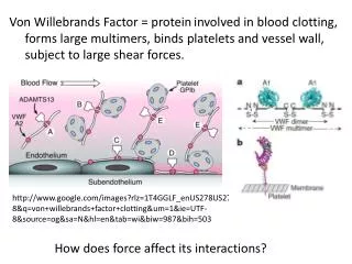
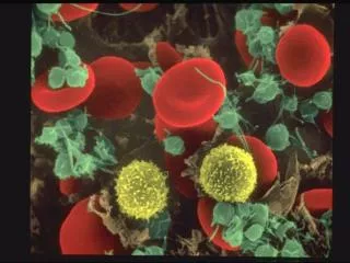
VON WILLEBRAND FACTOR. Large Adhesive Glycoprotein Produced by endothelial cells Polypeptide chain: 220,000 MW Base structure: Dimer; Can have as many as 20 linked dimers Multimers linked by disulfide bridges Synthesized in endothelial cells & megakaryocytes

E N D
VON WILLEBRAND FACTOR • Large Adhesive Glycoprotein • Produced by endothelial cells • Polypeptide chain: 220,000 MW • Base structure: Dimer; Can have as many as 20 linked dimers • Multimers linked by disulfide bridges • Synthesized in endothelial cells & megakaryocytes • Constitutive & stimulated secretion • Large multimers stored in Weibel-Palade bodies • Functions:1) Stabilizes Factor VIII2) Essential for platelet adhesion
Tissue factor • Under physiological conditions TF is expressed by cells not in contact with blood such as vascular smooth muscle, mesenchymal and epithelial cells including placental villous stromal cells.
Regulation of the Clotting Cascade • Almost every protease in the body has a corresponding protein inhibitor. • Serpins (serine protease inhibitors) • For thrombin this is the enzyme antithrombin III. During tissue damage, mast cells release heparin, a polysaccharide that enhances the activity of antithrombin III. However, it is not very effective against Fibrin bound thrombin, so its role is to limit thrombin activity away from the site of clotting.
Regulation (continued) • Thrombin initiates clot formation, but also activates Protein C. • This is the initial step in dismantling the blood clotting cascade. • Protein C is also a serine protease. Its targets are the non-enzymatic cofactors of the clotting cascade, factors V & VIII. • By deactivating these cofactors the growth of new clot material is slowed and eventually stopped. Genetic defects in Protein C activity result in thrombosis. A severe, recessive form of the disease results in neonatal death.
Anti-thrombin III & Heparin Protein C
Dissolving the Clot and Anticoagulants Tissue Plasminogen activator (TPA). Figure 16-14: Coagulation and fibrinolysis
Complement Activation • The Complement system is a complex cascade involving approximately 30 glycoproteins present in serum as well as cell surface receptors; • Activation of inflammation and immune related function; • Blood-materials interactions-protein adsorption;
Chronic Inflammation Macrophages produce great number of biologically active products • proteases • chemotactic factors • coagulation factors • growth promoting factors • cytokines • Growth factors (e.g. PDGF, FGF, TGF-b, IL-1, TNF, VEGF) are essential for: the growth of fibroblasts and blood vessels and the regeneration of epithelial cells • stimulate the production of a wide variety of cells • initiate cell migration and differentiation • tissue remodeling and wound healing
Fibroblast Play a Major Role in Wound Repair • Migration into the wound site • Integrin mediated cell-matrix attachment • Activated in response to PDGF, FGF and TGF-beta • Produce extracellular matrix • Produce VEGF-stimulate angiogenesis The combination of ECM, fibroblasts and new blood vessels is often referred to as granulation tissue. • Differentiation into Myofibroblasts-smooth muscle like phenotype-alpha-smooth muscle actin and myosin-like motor proteins that augment contractile force. • Contraction of Wound-Fibrous encapsulation • Disappear by apoptosis after wound closure
Granulation Tissue • the hallmark of early stage of healing (inflammation) • derives its name from the pink, soft granular appearance on the surface of healing wounds • may be seen as early as 3-5 days following implantation of a biomaterial • New small blood vessels are formed by budding or sprouting of preexisting vessels in a process known as “neovascularization” or “angiogenesis” • Angiogenesis involves proliferation, maturation, and organization of endothelial cells into capillary tubes
Angiogenesis-growth of new blood vessels • Under normal conditions, angiogenesis is quiescent in the adult human and stimulation of new blood vessels is thought to result from an alteration in the local balance of pro-angiogenic and anti-angiogenic growth factors.
Angiogenesis-growth of new blood vessels • Induced by low oxygen levels • Induced by low pH • Induced by elevation of cytokines • New blood vessels deliver oxygen, nutrients and inflammatory cells to the wound site that facilitates removal of debris. • Increase in oxygen enhances collagen synthesis so it is thought that angiogensis must precede extracellular matrix maturation and remodelling.
Angiogenic Factors • VEGF-vascular endothelial growth factor • Secreted by macrophages and fibroblasts • bFGF-basic fibroblastic growth factor • bFGF-elevated initially and after 48 hours decreases to baseline, whereas VEGF levels peak several days after injury
Myofibroblasts • Main cellular type in granulation tissue • Contain abundant stress filaments and smooth muscle like contractile machinery • Are interconnected by gap junctions • Main cellular type involved in extracellular matrix deposition;
Wound Contraction & Scarring • Late stage process • Cells at the wound site generate tractional and contractional forces on secreted matrix molecules to assist in wound closure. • The number of cells, the amount of matrix deposited and the force exerted determines whether the wound will close appropriately, as well as the amount of scar tissue produced-encapsulation tissue.
Mechanical Attachment of Cells • Key event in the process • May regulate whether the process occurs normally or in a pathologic manner. • Involves a variety of matrix components and may be thought of as consisting of a series of linked stages including: • Initial cell matrix contact • Recruitment of attachment sites to focal contact formation; • Cytoskeletal organization and spreading; • Cell-matrix tractional force generation and eventual cell contraction; • Matrix deformation (shortening)
Outcomes of Acute Inflammation • Complete resolution • Limited tissue injury or short lived inflammation • In tissue capable of regeneration • Removal of chemical mediators • Normalization of vascular permeability • Cessation of leukocyte emigration • lymph drainage clear edema, cells and debris • Scarring or fibrosis • Abscess formation • Progression to chronic inflammation
Chronic Inflammation Persistent inflammatory stimuli such as a foreign body or biomaterial lead to chronic inflammation: • chemical and physical properties of biomaterial • motion in the implant site • Confined to the implant site
Foreign Body Reaction consists of: • multinucleated foreign body giant cells • macrophages • fibroblasts • capillaries • Multinucleated foreign body giant cells form upon coalescence of macrophages
Foreign Body Giant cells • Fusion of 100s of macrophages and monocytes
FBR Depends on the Geometry and the Form of the Implant flat and smooth surfaces such as those found on breast prostheses; FBR is composed of a layer of macrophages one to two cells in thickness relatively rough surfaces such as those found on the outer surfaces of vascular prosthesis; FBR composed of multiple layers of macrophages and foreign body giant cells at the surface rough surfaces such as fabric-type materials; composed of macrophages and foreign body giant cells with varying degrees of granulation tissue
Foreign Body Reaction • FBR consisting mainly of macrophages and foreign body giant cells may persist at the tissue implant interface for the lifetime of the implant • FBR is surrounded by a fibrous tissue that isolates the implant from the local tissue environment • it is not known whether they remain activated releasing their lysosomal contents, or become quiescent.
Fibrous Encapsulation • End stage of healing response • Usually four or more weeks after implantation • A relatively acellular fibrous capsule • spindle shaped fibroblasts • small number of macrophages • Presence of neutrophils suggests persisting inflammatory challenge • Presence of macrophages suggests production of small particles by corrosion, depolymerization, dissolution or wear
Fibrous Encapsulation • Presence of lymphocytes suggests specific immune response • Thickness of the capsule depends on the chemical activity (rate of release) of the material: • metals which corrode freely • polymers with leachable constituents • Capsule thickness will increase with relative motion between the implant and the tissue • Shape of the implant: capsule will be thicker over sharp edges
Possible outcomes for the implant: • resorption: if the implant is resorbable then the implant site eventually resolves to a collapsed scar or, in the case of bone, may completely disappear • integration: very limited occurrence in practice; close approximation of normal host tissue to the implant without an intervening capsule (e.g. implantation of pure titanium in bone) • encapsulation: the most usual response