THROMBOPHLEBITIS
Definition.Histology of Vein.Anatomy of Vein.Thrombophlebitis. (pathogenisis)Varicose veins.Symptoms.Treatment.Complications.Prevention.. Contents. Thrombus : clot inside the vein.Phlebitis : inflammation of the vein.Thrombophlebitis: Inflammation of a vein that occurs when a blood clot


THROMBOPHLEBITIS
E N D
Presentation Transcript
1. By: Abdullah Al Moqawed
Abdullah Al Waznah
Abdulmajeed Al Majid
Abdulaziz Al Zaid THROMBOPHLEBITIS
2. Definition.
Histology of Vein.
Anatomy of Vein.
Thrombophlebitis. (pathogenisis)
Varicose veins.
Symptoms.
Treatment.
Complications.
Prevention. Contents
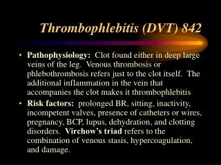
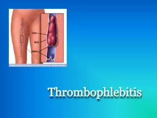
3. Thrombus : clot inside the vein.
Phlebitis : inflammation of the vein.
Thrombophlebitis: Inflammation of a vein that occurs when a blood clot forms. Definition
4. The blood vessels are made of three layers, called from the luminal side outward, the tunica intima, the tunica media and the tunica adventitia. These three layers are analogous to the endo-, myo- and epicardium, respectively.
The thickness of these three layers varies greatly depending upon the size and type of vessel (large, medium and small arteries and veins; capillaries).
Histology of Blood Vessels
5. The Tunica Intima:
Consists of an endothelium (present in all vessels) and any subendothelial connective tissue that may be present (highly variable depending on vessel). The endothelium of vessels entering or leaving the heart is continuous with that of the heart.
Histology of Blood Vessels (Cont.)
6. The Tunica Media:
Is the layer of concentrically-arranged smooth muscle, the autonomic control of which can alter the diameter of the vessel and affect the blood pressure.
The tunica media of arteries is larger than that of veins of similar size.
The tunica adventitia:
Is made chiefly of longitudinally arranged collagen fibers. It tends to be much larger in veins than arteries.
Histology of Blood Vessels (Cont.)
7. Veins function to return deoxygenated blood to the heart, and are essentially tubes that collapse when their lumens are not filled with blood.
The thick, outer-most layer of a vein is comprised of collagen, wrapped in bands of smooth muscle while the interior is lined with endothelial cells.
Most veins have one-way flaps called venous valves that prevent blood from back flowing and pooling in the lower extremities due to the effects of gravity. Anatomy of Venous System In the leg the long saphenous vein passes anterior to the medial malleolus at the ankle, up the medial aspect of the calf to behind the knee, then up the medial aspect of the thigh to join the common femoral vein in the groin at the saphenofemoral junction.
The short saphenous vein passes behind the lateral malleolus at the ankle and up the posterior aspect of the calf. It commonly joins the popliteal vein at the saphenopopliteal junction, which usually lies 2 cm above the posterior knee crease.
There are many intercommunications between the long and short saphenous systems and the venous anatomy of the leg is highly variable.
In the leg the long saphenous vein passes anterior to the medial malleolus at the ankle, up the medial aspect of the calf to behind the knee, then up the medial aspect of the thigh to join the common femoral vein in the groin at the saphenofemoral junction.
The short saphenous vein passes behind the lateral malleolus at the ankle and up the posterior aspect of the calf. It commonly joins the popliteal vein at the saphenopopliteal junction, which usually lies 2 cm above the posterior knee crease.
There are many intercommunications between the long and short saphenous systems and the venous anatomy of the leg is highly variable.
8. Anatomy of Venous System (Cont.)