Hemoglobin – 3D Structure
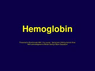
Hemoglobin Presented to Bioinformatic MN1 10 p course, Spring term 2006 by Amirah Khan With acknowlegment to Miriam Geörg & Björn Garpefjord. Hemoglobin – 3D Structure. Tetrameric complex a 2 b 2 Chain A = Chain C Chain B = Chain D 1 heme group per subunit/chain CATH Classification

Hemoglobin – 3D Structure
E N D
Presentation Transcript
HemoglobinPresented to Bioinformatic MN1 10 p course, Spring term 2006 by Amirah Khan With acknowlegment to Miriam Geörg & Björn Garpefjord
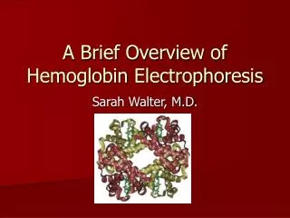
Hemoglobin – 3D Structure Tetrameric complex a2b2 Chain A = Chain C Chain B = Chain D 1 heme group per subunit/chain CATH Classification C Mainly Alpha (1) A Orthogonal Bundle T Globin-like H Globin
N-capping of helices • Helix: Ala53 – Ala71 • Removal of Ala53 • Replaced by Asp, negatively charged • Reduced dipole moment
N-capping of helices • Reduced dipole moment in helix • Indication of H-bond to Gly57 • More stable?
Reducing the Flexibility of the Main Chain • Stabilization of exposed loop by mutating Gly to Pro Mutation: G51 P
Stabilizing the Quartenary Structure • The hemoglobin tetramer consists of 2 ab dimers. • The interface between those 2 dimers is important for flexibility and funcionality. • Stabilization of the ab dimers by creation of disulfide-bonds between the subunits Mutations: a chain: Ala 123 Cys b chain: Val 33 Cys
Stabilizing the Hydrophobic Core • The main pocket of each chain is occupied by the heme group which is essential for function. • Mutations in these pockets might interfer with heme binding and thus oxygen transport.
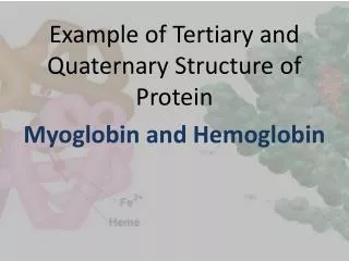
Increasing the Oxygen - Affinity • In nature there exist different forms of hemoglobin: • Adult hemoglobin (a2b2) • Fetal hemoglobin (a2g2) with higher oxygen affinity • Increase of Oxygen Affinity by mutating the aa’s close to the coordinative His 92 to the corresponding aa’s in fHb Coordinative Histidine
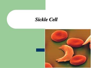
Stabilized Structure Industrial Applications? Conserved Structure Optimized by evolution!!! Flexibility of subunits needed for protein function Induced fit: conformational change after binding of first O2 leads to increased affinity for following O2 molecules Large pockets occupied by essential heme group Single aa exchange in Sickle Cell Anemia causes aggregation of hemoglobin Stabilization vs Flexibility