Download
1 / 34
340 likes | 622 Views
Chapter 7— The Pancreatic Islets focusing on insulin. Ch. 7-- Study Guide .

E N D
Ch. 7-- Study Guide Critically read (1) pages pp. 129-130 before Glucagon section; skip Glucagon section (pp. 130-134); (2) pages 134-143 (Insulin section) before Mechanism of insulin action subsection; skip Mechanism of insulin action subsection (p. 143). (3) pages 144-146 before Hormonal and neural control subsection Comprehend Terminology (the text in bold/italic) Study and understand the text and corresponding figures.
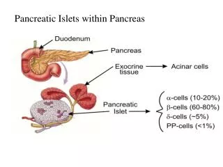
7.1. Introduction— Where is the pancreas?
§ Introduction • Principal pancreatic hormones: insulin and glucagon (both are _____ hormones) • Function of the insulin– an anabolic hormone that promotes CHO, fat, and protein storage • (Locations) Insulin’s target organs– skeletal muscle, liver, and adipose tissue • Diseases– • Diabetes mellitus type I– insufficient production • Diabetes mellitus type II—decreased end organ sensitivity to insulin
§ The pancreas and islets • Islets makes up about 2% of the pancreas • Blood circulation– by the pancreatic artery and drains into the portal vein toward the liver • The islets--highly vascular and each cell seems in directly contact with a capillary • (Nerve) The islets are innervated richly with sympathetic and parasympathetic fibers • Histology—Three major cell types: Fig. 7.1 + x • Beta cells (60% of the islet)– produce insulin • Alpha cells (30% of the islet)– produce glucagon • Delta cells (the least)—produce somatostatin
§ Biosynthesis and metabolism • Biosynthesis and secretion [in _____ cells of the ________ (what organ)]: Fig. 7.8 • (At ER) Preproinsulin becomes proinsulin– by cutting of the leader sequence; forming single chain proinsulin and disulfide bond formation • (at Golgi) packaging • (Forming mature insulin in granules)–converting into insulin (two chains) by cutting away the C (connecting) peptide • Metabolism (mainly in the liver)– half-life about 5 minutes; using receptor-mediated endosomic mechanism; also in kidneys, muscle, and others
(110 amino acids) A--at E. R. B--Golgi (51 a.a.) (31 a.a.) (110 amino acids) C--in secretory granules
7.4. Insulin—Physiological actions of insulin: A.Effects of insulin deficiency
§ signs of insulin deficiency (A) • Hyperglycemia– normal individuals (90 mg/dL of plasma); in diabetics: 300 to 400 mg/dL; testing diabetics by oral glucose tolerance tests (Fig. 7.9) • Glycosuria– excretion of glucose in urine; this is due to too much ___ in the blood & renal filtrate • Polyphagia/Polydipsia/Polyuria– • Excessive food consumption— due to increased in appetite to compensate for urinary lose of glucose • Excessive drinking– due to dehydration • Excessive urine– due to increased glucose in renal filtrate
§ signs of insulin deficiency (B) • Weight loss– • Underutilization of glucose by muscle/adipose tissue and overproduction of glucose by liver. These phenomena lead to hyperglycemia and items below. • due to reduced anabolic processes and increased catabolic processes – severe reduction in the ability to store glycogen, fat, and protein– marked wasting of muscle with depletion of body fat stores • This leads to lipemia (high lipid conc. In blood) • Increased fatty acids oxidation by the liver resulting in increased production of the ketone bodies (ketosis); plasma pH may become too low (acidotic coma and death possible)
§ Insulin’s effects • Target organs– adipose tissue, skeletal muscle, and liver • Physiology– • on above organs to defend and expand reserves of triglyceride, glycogen, and protein • Lower plasma glucose conc., amino acids, FFA etc. (If blood glucose too low – hypoglycemic coma) • How? (A) insulin increases glucose uptake by muscle and adipose tissue; (B) insulin decreases glucose output by liver; (C) by stimulating a.a. uptake by muscle; (D) by blocking FFA release from adipocytes
7.4. Insulin—Physiological actions of insulin: B.Effects on adipose tissue
§ Insulin on adipose tissue • Transport of glucose into adipocytes– via glucose transporters (GLUTs) • Accelerating glucose oxidation– increase availability of glycerol phosphate • Promoting FAA uptake from the lipoproteins of blood • Stimulating synthesis of triglycerides Fig. 7.10, 7.11, X
Reactions enhanced by insulin (green arrows) are 1-5. Reactions inhibited by insulin (dashed red arrows). In adipocyte
B A In adipocyte Confocal fluorescent microscope images of cultured mouse adipocytes that were transfected with GLUT4 linked to green fluorescent protein and then incubated in the absence (A) or presence (B) of insulin for 30 min.
7.4. Insulin—Physiological actions of insulin: C.Effects on muscle
§ Insulin on muscle • Promotes glucose transport into muscle– via glucose transporters (GLUTs); similar to that in adipocytes • Promotes glycogen storage– • Promotes protein synthesis Fig. 7.12, 7.13
7.4. Insulin—Physiological actions of insulin: D.Effects on liver
§ Insulin on liver • Promotes glycogen storage and reduces outflow of glucose – • In general, liver takes up glucose when the circulating conc. Is high and releases it when the blood level is low. • Movement of glucose is passive and depends on the glucose transporter. • Promotes fatty acid and protein synthesis • Substrates from myocytes and adipocytes influence hepatic effects Fig. 7.14
§ Regulation of insulin secretion • Insulin secretion is greatest right after eating – Fig. 7.20 • How does it work? • Coordination of insulin secretion with nutritional state is via direct monitoring of metabolites, hormones, and neural signals by beta cells. • Glucose– is the most important regulator of insulin secretion; however, beta cells do not have specific receptors for glucose!!! • Other metabolites– a.a. and fatty acids are important stimuli for insulin secretion