Download
1 / 142
1.42k likes | 1.44k Views
Explore the anatomy of the heart, vascular system, cell communication, and electrical activity of the heart. Learn about cardiomyocytes, heart wall layers, and heart rhythm patterns. Textbooks used: Human Physiology – An Integrated Approach, Human Physiology, Functions of the CVS.

E N D
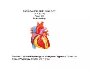
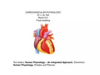
CARDIOVASCULAR PHYSIOLOGY Dr J. du Toit Room 511 Fisan building Text books: Human Physiology – An Integrated Approach. Silverthorn. Human Physiology. Rhodes and Pflanzer.
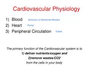
Functions of the CVS • Transport of: 1) nutrients and water and, 2) gases DS Lungs • Cells Liver Cells • Kidneys • Cell cell communication - hormones • Transport – fatty acids from adipose tissue & glucose from the liver • Transport WBC & antibodies • NB in temperature regulation
Anatomy of the Heart • Size of fist • Between lungs - base under sternum and apex on diaphram Epi- en pericardium – fibrous and serous tissue WallMyocardium - contractile cells Endocardium – endothelium –continuous with blood vessel endothelium • 2 Atria • 4 Chambers 2 Ventricles
Right – Tricuspid valve (3-leaflets) • • Atria & ventricles separated by AV valves Cordae tendinae en papillary muscles • Left – Bicuspid – Mitral valve (2 leaflets) • Ventricles of pulmonary artery and aorta – separated by – semilunar valves: Aortic valve and • Pulmonary valve • Arteries oxygenated blood - red VASCULAR SYSTEM Veins deoxygenated blood - blue
The Heart wall a) Epicardium/Pericardium: 2 layers nl. i) Outside fibrous pericardium ii) Inside serous pericardium Functions: 1) Prevents excess stretching of heart 2) Provides smooth, lubricated outside surface b) Myocardium: Contractile part of the heart wall. c) Endocardium: Connective tissue attaches the myocardium to the endothelium. The latter provides smooth surface and prevents clotting.
Properties of the cardiomyocyte: • Is striated • Contains one or more nuclei • Cells are branched • More mitochondria than skeletal muscle • Contains tight junctions and gap junctions. • 2 Types of cardiomiocytes: • 1) normal cardiomyocytes • 2) pacemaker and conducting cardiomyocytes ELECTRICAL ACTIVITY OF THE HEART Structure of the cardiomyocyte
AP of Skeletal and Heart muscle • Depolarisation due to Na+ influx • Repolarisation due to K+ efflux • Heart muscle AP – has plato due to Ca2+ influx longer AP
Importance of the Refractory Period • Prevents tetanus • Ensures diastolic relaxation
SA node • wall of the RA near superior vena cava. • primary pacemaker at rest = 70bpm. • Parasympathetic: ACh - heart rate • Sympathetic: adren. & nor-adren. - heart rate and contractile force • Sensitive to – temp., stretch, touch and chem. stimulation • AV node • Bottom wall of the RA - interatrial septum • Firing frequency: 40-60bpm 1) Delays heart impulse: 0.1 sec complete ventricular filling 2) Delays frequency of impuls propagation
AV bundle • From the AV-node to interventricular septum. • Right bundle branch – right of the septum to the apex of the heart • Left bundle branch – posterior/inferior branch • anterior/superior branch • functional link between atria and ventricles • Purkinje fibres • branches of the left and right bundle branch • impulse propagation to contractile cells in ventricle
AP – Origin and Propagation SA-node propagation speed fast (1 m/sec) AV-node propagation speed slow (0.05-0.1 m/sec) no direct conduction from atria to ventricle muscle – Fibrous plate/sheath Bundle of His propagation speed fast Purkinje system propagation speed fast (2 m/sec) Ventricular contractile cells
Wave of depolarization over heart creates a potential difference - dipole • Dipole (hart) surrounded by conductor (elektroliete & water) • Elektrodes on surface attached to galvanometer – measures potential differences ECG – measures electrical changes in heart
ECG • The sum of all the potentials that are created by the cells of the heart at any given moment • Each component of the ECG reflects a de- or repolarisation of a part of the heart can associate parts of the ECG with parts of the cardiac cycle. • Clinical application of the ECG • Determination of: • HR, heart rhythm • Presence of hypertrophy or atrophy • Abnormal conduction paterns • The cardiac axis (electrical axis) • Normal heart rhythm • Sinus rhythm – bradycardia or tagycardia
ECG Leads • Position of the electrodes = leads • 6 peripheral leads and 6 precordial leads • a) 3 bipolar limb leads (standard leads) measure the potential differences between 2 points (Einthoven triangle) • b) 9 unipolar leads measure the potential at a point on the body • •3 unipolar limb leads: aVR, aVL en aVF • • 6 unipolar chest leads: V1-V6
Normal heart rhythm • Sinus rhythm: • Sinus tachycardia: > 100 bpm • Causes: exercise, emotional excitement, heart failure, fever, anemia. • Sinus bradycardia: < 60 bpm • Causes: long term exercise, hypothyroidism. • Sinus arrhythmias: irregular firing of the SA-node (fast and slow beats) • Ectopic heart beats: the impulse that causes heart contraction originates outside • the SA-node and causes extrasystoles.
The QRS complex may be abnormally large (ventricular hypertrophy) or • abnormally small (ventricular atrophy). • The QRS complex does not follow the P-wave. Sometimes several P-waves • followed by the QRS-complex. Causes, heart block. The impulse is not always conducted through the AV-node. • The Q-wave is enlarged, abnormal QRS-complex, ST-segment is elevated above baseline and inverted T-waves are indicative of necrotic heart muscle. Therefore MI.
Abnormal ECG Heart defect ECG Defect Ventricular hypertrophy QRS complex with high amplitude Ventricular atrophy QRS complex with low amplitude Ischaemia and infarction Abnormal QRS complex, ST segment elevated, T-wave inverted. Bundle branch block wide QRS complex due to delayed conduction Heart block P wave not followed by QRS complex 1st degree delayed QRS complex 2nddegree absent QRS complex 3rd degree total AV dissosiasion VT Abnormal QRS complex, no P wave VF No QRS complex distinguishable
Heart sounds 1st heart sound – during ventricular systole – low tone – closing of AV valves 2nd heart sound – end of ventricular systole – sharp with high tone – closing of the semilunar valves. 3rd heart sounds – end of systole - AV valves open – blood flows through 4th heart sounds – artial systole – vibrasion of the ventricular wall