Download
1 / 94
940 likes | 988 Views
Explore the organization and functions of the Nervous System, including the Central and Peripheral Nervous Systems, Motor Division, and Autonomic Nervous System subdivisions. Learn about neurons, reflex arcs, and ANS control of physiological functions. Dive into Neuroglia cells and their roles in supporting nerve cells. Discover the intricacies of nerve impulse transmission, neuron structure, and functional neuron classification.

E N D
Organization of the Nervous System • Central Nervous System (CNS) – • Brain and spinal cord • Peripheral Nervous System (PNS) – • Made up of nerves that attach to CNS
P e r i p h e r a l N e r v o u s S y s t e m S k e l e t a l A u t o n o m i c ( S o m a t i c ) S y m p a t h e t i c P a r a s y m p a t h e t i c Peripheral Nervous System
Functional Subdivisions of the PNS Name "A"! • PNS contains 2 functional subdivisions • Sensory (afferent) nerves conduct sensory impulses to CNS from receptors in organs and tissues • Motor (efferent) nerves conduct impulses away from CNS to muscle effectors and glands Now "C"? What's "B"?
Subdivisions of Motor Division (PNS) • Somatic Nervous System (SNS) – • Controls voluntary and involuntary skeletal muscle contractions • Involuntary skeletal contraction – reflex • Single neuron system • Autonomic Nervous System (ANS) – • Controls activity of smooth and cardiac muscles • 2 neuron system • 1st: from CNS to ganglion • 2nd: from ganglion to effector
Subdivisions of ANS • Sympathetic division – • Fight or flight • Parasympathetic division – • Regulates resting functions • Ex., digesting food Color-code your picture of the NERVOUS SYSTEM!
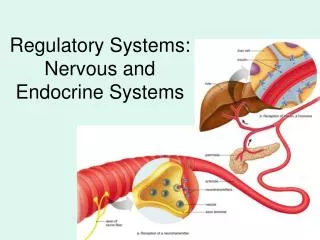
CENTRAL NERVOUS SYSTEM Brain Dilates pupil Stimulates salivation Salivary glands Relaxes bronchi Spinal cord Lungs Accelerates heartbeat Heart Inhibits activity Stomach Pancreas Stimulates glucose Liver Adrenal gland Secretion of adrenaline, nonadrenaline Kidney Relaxes bladder Sympathetic ganglia Stimulates ejaculation in male Sympathetic Inhibits • “ Fight or flight” response • Release adrenaline and noradrenaline • Increases heart rate and blood pressure • Increases blood flow to skeletal muscles • Inhibits digestive functions
CENTRAL NERVOUS SYSTEM Brain Contracts pupil Stimulates salivation Constricts bronchi Spinal cord Slows heartbeat Stimulates activity Stimulates gallbladder Gallbladder Contracts bladder Stimulates erection of sex organs Parasympathetic • “ Rest and digest ” system • Calms body to conserve and maintain energy • Lowers heartbeat, breathing rate, blood pressure
Autonomic nervous system controls physiological arousal Sympathetic division (arousing) Parasympathetic division (calming) Pupils dilate EYES Pupils contract Decreases SALIVATION Increases Perspires SKIN Dries Increases RESPIRATION Decreases Accelerates HEART Slows Inhibits DIGESTION Activates Secrete stress hormones ADRENAL GLANDS Decrease secretion of stress hormones Summary of ANS differences
Nervous Tissue • Made up primarily of 2 types of cells: • Neurons • Supporting cells • CNS - neuroglia (glial cells) • Astrocytes, microglia, ependymal cells, oligodendrocytes • PNS – satellite cells, Schwann cells
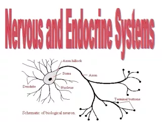
Neurons • Made up of a cell bodies, dendrites, and axons • Cell body (or soma) • Contains large nucleus and Nissl bodies (Rough E.R.), primary site of protein synthesis • Most located within CNS where they are protected by hard bones of skull and vertebral column • Collection in CNS is called “nuclei” whereas in PNS they are called “ganglia”
More on Neuron Structure • Dendrites • Short, highly branched signal receptive regions of nerve cell • Convey incoming message toward the cell bodies • Each nerve cell (neuron) has only one axon • Impulse generating and conducting region of neuron
Myelin Sheath of the Axon • Whitish, fatty protein layer • Serves to protect and electrically insulate axon • Increases the speed of transmission of nerve impulses (up to 150 times faster) • Only associated with axons, not dendrites
REVIEW:Functional Classification • Neurons are grouped according to the direction in which the nerve impulse travels relative to the CNS • Based on this there are sensory, motor, and association neurons • Sensory – transmit impulses from skin or other organs toward CNS • Motor – carry impulses away from CNS to effector organs • Association (interneurons) – lie between motor and sensory neurons
Label your Neuron Neuron Video neurilemma
Neuroglia (CNS) • CNS has 4 different types of supporting cells (neuroglia) • Abundant and diverse • Limited knowledge of each function due to difficulty in isolating individual cells • Ependymal cells • Line central cavities of brain and spinal cord • Beating of cilia help to circulate the cerebrospinal fluid (CSF) • CSF: protection of brain and transport of vital nutrients and waste • Astrocytes • Attach neurons to capillaries, linking them to nutrient supply
Neuroglia CONT…. • Oligodendrocytes • Branches wrap around neuron fibers, forming an insulating covering called a myelin sheath • Microglia • Monitor health of nerve cells • If foreign invader detected or cell damaged, many microglia head to the area, transform into macrophages, and protect the CNS by phagocytizing the microorganisms or neuronal debris
Label your Synapses Diagram Use pages 392 and 406 in the AP book • HINTS FOR YOU: = Sodium ions = Potassium ions = Calcium ions = synaptic vesicles w/ neurotransmitters = chemical gated channels
Action potential • http://highered.mcgraw-hill.com/sites/0072495855/student_view0/chapter14/animation__the_nerve_impulse.html
ANIMATION OF CHEMICAL SYNAPSES • http://highered.mcgraw-hill.com/sites/0072495855/student_view0/chapter14/animation__chemical_synapse__quiz_1_.html Animation of ENTIRE SYNAPTIC PROCESS with ACTION POTENTIAL
Sodium/Potassium Pump • http://highered.mcgraw-hill.com/sites/0072495855/student_view0/chapter2/animation__how_the_sodium_potassium_pump_works.html
Action Potential Graph • Know what happens at each part of the graph with relationship to the pre-synaptic and post-synaptic activities. • Look at the HANDOUT with the graph!!
Action Potentials • Also known as “Nerve Impulses” • Self-regenerating wave of electrochemical activity that allows neurons to carry a signal over a distance (“game of telephone”) • Pulse-like waves of voltage that travel along several types of cell membranes • http://outreach.mcb.harvard.edu/animations/ actionpotential.swf
Action Potentials • Initiation/Resting Stage: • Some K+ channels are open: K+ diffusion occurring • Initiated by stimulus above a certain intensity or threshold(~-70mV – resting potential) • Could be a pin prick, light, heat, sound or an electrical disturbance in another part of the neuron (“telephone call”) • Electrical signal rises from changes in permeability of the neuron’s axon membranes to specific ions (Na+&K+)
Action Potentials Depolarization (Rising Phase) • K+ Channelgates are closed • Stimulus causes gate in the Na+ Channel to open • High concentration of Na+ outside, Na+ diffuses into neuron • Electrical potential changes to ~ +40 mV. Repolarization (Falling Phase) • Depolarization causes K+ Channelgate to immediately open & Na+ Channel close • K+ diffuses out of neuron • Reestablishment of initial electrical potential of ~-60 mV.
Action Potentials Refractory Period (Recovery Phase) • Na+ & K+ Channels cannot be opened by a stimulus • Na+/K+ Pump actively (ATP) pumps Na+ out of & K+ into neuron • Reestablishment of ion distribution of resting neuron • This AP acts as stimulus to neighboring proteins & initiates AP in another part of neuron • Wave of APs travel from dendrites to axon terminals • At axon terminal, electrical impulse is converted to a chemical signal (neurotransmitter)
mV= membrane voltage (within cell) Action Potentials
Complete the "Analyzing Changes During a Nerve Impulse" wkst So let's recap what we've talked about... Per. 2 start, Fri Per. 4, WU go over handout http://bcs.whfreeman.com/thelifewire/content/chp44/4402002.html
Action Potentials • Getting the message across (the synapse)? • http://www.mind.ilstu.edu/curriculum/neurons_intro/neurons_intro.php • At axon terminal, chemical signal (NT) crosses synapse between adjacent neurons • Starts AP on this neuron • This activates Ca2+ channel to open • Ca2+ diffuses into neuron • Causes NT vesicles to move to end & fuse with cell membrane • Through exocytosis, NTs are released into synapse • NTs diffuse across synapse & bind to NT receptors on another neuron • Causes Na+ channels to open & AP is initiated in next neuron
How does the message get passed along? • Information from one neuron flows to another neuron across a synapse… • a small gap separating neurons that consists of: • a presynaptic ending that contains neurotransmitters, mitochondria & other organelles, • a postsynaptic ending that contains receptor sites for neurotransmitters & • a synaptic cleft or space between the presynaptic & postsynaptic endings. Label & color-code the back of your Neuron wkst!
Let’s Review…How do neurons communicate? • What is another name for the nerve impulse that travels from the axon hillock through the axon to the axon terminal? • What are the 4 main phases of an Action Potential? • What happens in the Rising Phase? • Falling Phase? • Recovery Phase? • Resting Potential? • What ion entering the neuron instigates the movement of synaptic vesicles to the cell membrane? • What do the vesicles release into the synapse? • What happens after the NTs are released into the synapse?
Brain Spinal Cord Central Nervous System • Brain and Spinal Cord
The Human Brain • Average adult male’s brain weighs approximately 1.6 kg (~3.5 lbs) • Average female’s: 1.42kg • According to body weight, though…they are relatively equal in size
Corpus Callosum Right Hemisphere Left Hemisphere Brain has 2 Hemispheres • Left & Right sides are separate • Corpus Callosum : major pathway between hemispheres • Some functions are ‘lateralized’ • Language, numbers on left • Color and music on right • Lateralization is never 100%
Medial surface of right hemisphere Corpus Callosum Corpus Callosum • Major ( but not only) pathway between sides • Connects comparable structures on each side • Permits data received on one side to be processed in both hemispheres • Aids motor coordination of left and right side
Motor Cortex Somatosensory Cortex Contralateral Motor Control • Movements controlled by motor area • Right hemisphere controls left side of body • Left hemisphere controls right side • Motor nerves cross sides in spinal cord
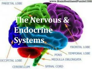
Frontal Parietal Occipital Temporal Each hemisphere is divided into 4 lobes Cerebellum
Protection of the Brain - Meninges • Cover and protect CNS • Protect blood vessels and enclose venous sinuses • Contain cerebrospinal fluid • Form divisions within the skull
Cerebral White Matter • Deep to the gray matter of cortex • Aids communication between cerebral areas and between cerebral cortex and lower CNS • Spinal cord has just the opposite type of matter http://www.brainexplorer.org/brain-images/white_matter.jpg
Frontal Lobe Working Memory Motor Cortex Motor Cortex Motor Cortex Broca’s Area Frontal Lobe • Contains primary motor cortex • No direct sensory input • Important planning and sequencing areas • Broca’s area for speech • Prefrontal area for working memory
Occipital Lobe • Input from Optic nerve • Contains primary visual cortex • most is on surface inside central fissure • Outputs to parietal and temporal lobes Occipital Lobe Visual Lobe
Auditory Cortex Temporal Lobe Temporal Lobe Temporal Lobe • Contains primary auditory cortex • Inputs are auditory, visual patterns • speech recognition • face recognition • word recognition • memory formation • Outputs to limbic System, basal Ganglia, and brainstem
Somatosensory Cortex Parietal Lobe Parietal Lobe • borders visual & auditory cortex • Inputs from multiple senses • Outputs to Frontal lobe • hand-eye coordination • eye movements • attention
Regions & Organization • Cerebral Hemispheres – • Has outer cortex of gray matter (neural bodies) • Diencephalon – • Thalamus, hypothalamus, and epithalamus • Brain Stem – • Midbrain, pons, medulla • Cerebellum – • Has outer cortex of gray matter
Label the worksheet on the brain!! • I will place the worksheet under the document camera for you to view!!
Cerebral Hemispheres • Approx. 83% of total brain mass • Covered with elevations called gyri and shallow grooves called sulci • Deeper grooves, called fissures, separate major regions of the brain