Download
1 / 10
100 likes | 243 Views
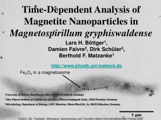
Time-Dependent Analysis of Magnetite Nanoparticles in Magnetospirillum gryphiswaldense. Lars H. Böttger 1 , Damien Faivre 2 , Dirk Schüler 3 , Berthold F. Matzanke 1 http://www.physik.uni-luebeck.de. Fe 3 O 4 in a magnetosome.

E N D
Time-Dependent Analysis of Magnetite Nanoparticles in Magnetospirillum gryphiswaldense Lars H. Böttger1, Damien Faivre2, Dirk Schüler3, Berthold F. Matzanke1 http://www.physik.uni-luebeck.de Fe3O4 in a magnetosome 1 University of Lübeck, Ratzeburger Allee 160, 23538 Lübeck, Germany 2Max Planck Institute of Colloids and Interfaces,Wissenschaftspark Golm, 14424 Potsdam, Germany 3 Microbiology, Department of Biology, LMU München, Maria-Ward-Str. 1a, 80638 München, Germany
In recent years magnetic material has been found in several species, such as termites [1], newts [2], pigeons [3], sea turtles [4] or magnetotactic bacteria [5]. In magnetotactic bacteria, magnetite crystals are surrounded by a lipid bilayer membrane, termed magnetosome. The magnetosome membrane is derived from the cytoplasmic membrane. The magnetosomes are aligned as a chain within a filamentous structure of the cytoplasm [6, 7]. Together with an oxygen sensing system, the magnetosome chain serves as a navigational device. Optimum growth conditions are found in an environment of low pO2 which is found in lower levels of ponds, lakes and rivers. Under microoxic conditions, tremendous amounts of iron are transported into the magnetotactic bacterium Magnetospirillum gryphiswaldenseand are metabolized (up to 4% of the dry weight [8]). Up to 100 cubooctahedral magnetite (Fe3O4) crystals per cell are formed biosynthetically. The unique crystalline and magnetic properties of magnetosomes have brought them into the focus of multidisciplinary interest because they are used in biotechnological applications [9] or as biomarkers for life on Mars [10].
Controlled growth in a fermenter • Fermenters allow control of several • parameters: • Temperature • pH • O2-partial pressure • Growth of magnetosomes without biomass production Figure 1: Typical fermenter setup To investigate the mechanism of magnetite-, magnetosome and magnetosome chain formation, the bacteria were grown in a fermenter under conditions of iron starvation. After harvesting the cells and reincubating them in a 57Fe-rich and C-poor medium, biosynthesis of magnetite is induced and a time-resolved analysis of the iron metabolite pattern can be achieved, which is not blurred by other metabolic processes. At defined times, cells were harvested, spun down in a centrifuge, washed and approximately 108 cells were transferred to Mössbauer sample holders.
Typical Mössbauer spectrum of Magnetospirillum with filled iron pools Figure 2: The spectrum is composed of 4 subspectra: two sextets representing magnetite A and B sites (1:2 ratio), a Fe(II) high-spin doublet and a ferritin-like Fe(III) doublet.
Mössbauer spectra of time dependent 57Fe-accumulation and iron metabolization Figure 3: Series of Mössbauer spectra taken of cells after the medium was incubated with57Fe-citrate. The magnetite contribution increases continuously during the accumulation period. Fe(II) remains at the same abolute levels and the ferritin-like component is growing slowly. The hyperfine field of the magnetite components increase for 155 min after 57Fe induction, but show a decrease of about 0.6 T afterwards. The red line is for comparision.
Chain formation time, determined by Bhf It has been shown by TEM that small magnetosomes containing a small magnetite nucleus are formed initially. Their iron content is growing in a time-dependent fashion. In a second step magnetosome chains are assembled. Chain formation has been analyzed by 3 independent methods: TEM, spectrophotometry and by analysis of the time-dependent changes of Bhf. The hyperfine field Bhf of an isolated magnetosome nanoparticle can be described by [11]: Bd is the contribution of the demagnetizing field. B0 is the value of hyperfine field that one would find in a macrocrystal.The cosine-term represents the thermal average of the cosine of the angle between the easy axis of magnetization and the actual magnetization. The cosine-termin the low temperature limit mainly depends on the term kBT/2V with kB representing the Boltzmann constant, T the temperature, the anisotropic energy constant and V the particle volume. The volume appears as time dependent term because of the ongoing growth of the magnetosomes/magnetite crystals. Therefore, the constants can be combined to a fit factor b and to the inverse of the time t.
Chain formation time, determined by Bhf As soon as the nanoparticles are arranged in a chain of magnetosomes (see figure 6) the demagnetizing field vanishes. Using an integrated Gaussian distribution G(t) describing the fraction of magnetic cells in a sample, one can describe the observed hyperfine field by: The term µ represents the transition time after which half of the bacteria have become magnetic, and the term represents the half width of the Gaussian distribution. A least square fit of magnetite A and B hyperfine fields yields the factor b, the curves shown in figure 4 and a transition time of µ = 175 min. The fit factor b allows to calculate the volume of particles and hence their diameter (assuming a sphere, see figure 5): A TEM series of the growth kinetics confirms both, the time of chain formation and the size of the magnetosome-bound magnetite crystals (see figure 6).
Chain formation time of magnetite nanoparticles determined by Bhf Figure 4 Fit of the hyperfine fields using a Gaussian distribution for magnetic cells. From this fit the chain formation time of 175 min was derived Size of magnetite nano-particles determined by Bhf Figure 5: Diameters of nanoparticles calculated from Mössbauer Bhf-data (solid lines) in comparison to diameters determined by TEM data (black stars)
0min 55min 100min 220min 340min TEM of the induction series Figure 6: Control of the chain formation in TEM: after 55 and 100 min isolated magnetosoms are visible. After 220 min most of the magnetosomes have been arranged in chain fragments. In the sample after 340 min the chain was fully formed. The arrows and ellipses emphasize the position of magnetosomes.
[1] Maher, Proc. R. Soc. Lond. B (1998) 265, 733-737 [2] Deutschlander, Borland and Phillips, Nature (1999) 400, 324-325 [3] Winklhofer, Holtkamp-Rötzler, Hanzlik, Fleissner and Petersen, Eur. J. Mineral. (2001) 13, 659-669 [4] Lohmann, Cain, Dodge and Lohmann, Science (2001) 294, 364-366 [5] Blakemore, Science (1975) 190, 377-379 [6] Komeili, Li, Newman and Jensen, Science 311,242-245 (2006) [7] Scheffel, Gruska, Faivre, Linaroudis, Plitzko and Schüler, Nature 440,110-114 (2006) [8] Lang, Schüler and Faivre, Macromol. Biosci. (2007) 7, 144-151 [9] McKay, Gibson, Thomas-Keprta, Vali, Romanek, Clemett, Chilier, Maechling and Zare, Science (1996) 273, 924-930 [10] Schüler and Bauerlein, J. Bacteriol. (1998) 180, 159-162 [11] Mørup, Dumesic and Topsøe,Magnetic microcrystals, in Applications of Mössbauer Spectroscopy, Cohen, 1980