Scanning Electron Microscopy
Scanning Electron Microscopy. 18 th January 2010. Topography The surface features of an object or "how it looks", its texture; direct relation between these features and materials properties Morphology The shape and size of the particles making up the object; direct

Scanning Electron Microscopy
E N D
Presentation Transcript
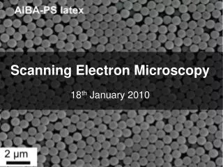
Scanning Electron Microscopy 18th January 2010
Topography The surface features of an object or "how it looks", its texture; direct relation between these features and materials properties Morphology The shape and size of the particles making up the object; direct relation between these structures and materials properties Composition The elements and compounds that the object is composed of and the relative amounts of them; direct relationship between composition and materials properties Crystallographic Information How the atoms are arranged in the object; direct relation between these arrangements and material properties
Why use it? Advantages Image bulk materials Large depth of field High resolution <10nm Quick and easy sample preparation (Only require sample to be conductive) More control in the degree of magnification etc Disadvantages Sputter coating can disturb delicate samples Lower resolution than TEM Expensive Samples must be dry
How it works! www.purdue.edu/REM/rs/sem.htm
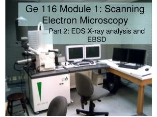
What’s available? FEI Sirion FEG : Sorby Centre 09:00 - 11:00 Via booking sheet 11:00 -13.00 13:00 -15:00 15.00 -17:00 £40 per hour SEM free for Tony Ryan's students: but only until October, Philips XL-20 : Dep. of Bio. Science 09:30-10:30 Via booking webpage 11:00-12:30 13:30-15:30 16:00-17:30 £19/£38 per hour dependent on the level of support required Prices will be increasing probably by around 10% shortly www.jeol.com/Default.aspx?TabId=115
Useful Contacts - Sorby Sorby centre: http://www.shef.ac.uk/materials/research/centres/sorby/ Sorby centre Manager Prof. W. M. Rainforth m.rainforth@sheffield.ac.uk Senior Experimental Officer Dr P Korgul p.korgul@sheffield.ac.uk Technical Staff Dr P Zeng p.zeng@sheffield.ac.uk (ESEM) Dr C Shields c.shields@sheffield.ac.ukext. 25519 (Main contact for SEM queries & training)
Useful contacts – Dep. Bio. Science Biomedical Science/Molecular Biology Electron Microscopy Suite: http://www.shef.ac.uk/bms/about/facilities/bclem/ Academic Director: Professor Per Bullough (p.bullough@sheffield.ac.uk) Molecular Biology Service Provider and Laboratory Manager: Dr Svetomir Tzokov (s.tzokov@sheffield.ac.uk) Biomedical Science Service Provider: Chris Hill (c.j.hill@sheffield.ac.uk) ext. 24674
Getting started - Sorby • Download access and registration form from Sorby website http://www.shef.ac.uk/materials/research/centres/sorby/ • Complete form with as much information as possible about what you want to do • Until October: • Financial Statement PAGE 2 • My research is free of charge (pre-agreed) • Get supervisor's signature • Arrange a time to for Mark Rainforth to sign form • Take signed form to Cathy Shields to arrange training All fields need to be answered and it needs to be signed by the supervisor and by Mark Rainforth before training sessions can be planned
Getting started – Dep. Bio. Sci • Contact staff • To arrange an initial consultation with regard to project • (Chris Hill is suggested SEM contact) • Invoices issued monthly • Send your SAP grant code to Chris Hill/Svetomir Tzodov, or pass invoices to finance administrator for payment and advise them to issue an internal trade number for your payment • Bookings made via website • Code of practice listed on website
Sample preparation • Good Specimen • Good conductor (gold coated if necessary – usually the case for polymers) • Must not degrade under the electron beam • Must not be hydrated • The sample should be held in a vacuum prior to going in the microscope. • Procedure • Dry sample, securely attach sample to SEM stub using carbon adhesive • Sputter coat with gold • SEM images • Save images on CD NOT USB – for Sorby centre
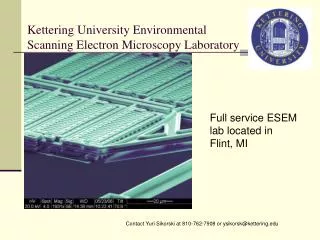
ESEM • Contact for ESEM is Peng Zeng email: p.zeng@sheffield.ac.uk • Doesn’t have to view samples in a vacuum. These images should be seen in a low pressure, wet environment. • No need for a conductive coating to be applied to sample