Download
1 / 19
210 likes | 455 Views
Cellular imaging at 3 T: Detecting Cells in Inflammation using Active Labeling with Super paramagnetic Iron oxide. Azhar Hosein Faraz Medical Biophysics Western University Robarts Research Institute Under supervision of: Dr. Paula Foster. 4 th April 2012.

E N D
Cellular imaging at 3 T: Detecting Cells in Inflammation using Active Labeling with Super paramagnetic Iron oxide AzharHosein Faraz Medical Biophysics Western University Robarts Research Institute Under supervision of: Dr. Paula Foster 4th April 2012
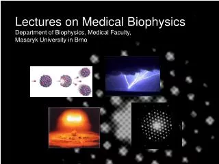
Immune Response and Inflammation • Tissue injury caused by bacteria, trauma, chemicals, or any other phenomenon –Inflammation. • Walling- off • Within minutes after inflammation occurs macrophages already present in the tissues (microglia, Kupffer cells,..) begins their phagocytic action. • The second line of defence within the first hours of inflammation begins are the large number of neutrophils that invade the inflamed area.
Imaging Immune Cells with MRI • In Vivo Labeling of Cells with Iron Particles Iron oxide contrast agent Labeled cells Intravenous (i.v) administration Phagocytosis
Iron oxide-based MRI contrast agents Iron-labeled cells In gel Effect of iron is to cause signal loss in MRI images Adapted from Modo, M. et al. Mol. Imag, 2004
Aim of study To detect inflammatory cells in the mouse brain by in vivo 3T MRI in a model of neuroinflammation.
Normal Healthy Mouse (C57/Bl6, n=2) Methods MRI Pre-Scan of body Iron Oxide (Fe) injected Intravenously (IV) to Mice • Fe • Bone marrow • Liver • Spleen Monocytes MRI Body 24 hours Post-injection (Fe) Inject mice with : LPS (lipopolysaccharide) 48 hours Post-injection (Fe) • Model of Neuroinflammation • Neurotoxic • Over activation of microglia MRI Body 6 days Post-injection (Fe) MRI Brain 9 days Post-injection (Fe)
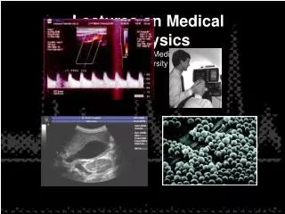
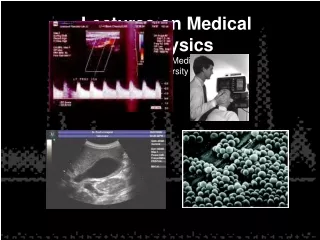
Imaging Cells (3T cellular MRI) at Robarts Custom-built high performance gradient • Only lab in the world • research imaging experiments at clinical magnetic strength • pulse sequence known as,bSSFP. 3T clinical system Solenoid radio frequency coil
Image Analysis: Body Images • Measuring mean signal intensity of different image slices in Liver, Bone marrow, and Spleen. • Standard deviation (SD) of Noise present in each slice. • SNR Signal to Noise ratio Mean signal intensity / SD of Noise Present in each scan slice
Image Analysis: BrainImages • No. of Voids present in Brain MRI scan • Mean signal intensity of each discrete region of signal void • Fractional signal loss in % = • Clear canvas software used for analyze of MRI scan images.
Whole Mouse Body BSSFP Images Before Iron Injection Tail Head Sagittal view of mouse
Post Injection Iron • In vivo labelling of liver, spleen and bone marrow macrophages
Results: SNR • Liver > Spleen > Bone marrow
MRI Brain • Voids present in brain
Results: FSL • FSL is related to the amount of Iron in discrete reigns of signal void. • Can be related to the number of iron-labeled cells.
SUMMARY • Changes in SNR suggests that cells take up iron in the liver, spleen and bone marrow - the numbers of iron-labeled cells is different for each organ and varies in mice. • This work indicates that pre-labeling immunecells with iron allows us to track their involvement in inflammation in the brain • This study has been done for first time
Future directions • To prove that signal loss in the brain is due to the accumulation of immune cells • To determine which kind of cells are presenting in brain, using histology and experiments with transgenic mice. • To obtain body images over a prolonged time period, to better understand the time course of cell uptake and retention of iron.
Acknowledgment • Supervisor : Dr. Paula Foster • Research funding : MS society • Thanking JonatanSnir, for imaging