Download
1 / 45
450 likes | 645 Views
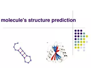
molecule's structure prediction. RNA RNA folding Dynamic programming for RNA secondary structure prediction Protein Secondary Structure Prediction Homology Modeling Protein Threading ab-initio. Outline. RNA Basics. 3 Hydrogen Bonds – more stable. 2 Hydrogen Bonds. RNA bases A,C,G,U

E N D
RNA RNA folding Dynamic programming for RNA secondary structure prediction Protein Secondary Structure Prediction Homology Modeling Protein Threading ab-initio Outline
RNA Basics 3 Hydrogen Bonds – more stable 2 Hydrogen Bonds • RNA bases A,C,G,U • Canonical Base Pairs • A-U • G-C • G-U “wobble” pairing • Bases can only pair with one other base. Image: http://www.bioalgorithms.info/
RNA Secondary Structure Pseudoknot Stem Interior Loop Single-Stranded Bulge Loop Junction (Multiloop) Hairpin loop Image– Wuchty
RNA secondary structure representation Circular representation: Bacillus Subtilis RNase P RNA
RNA secondary structure representation DotPlot representation of the same Bacillus Subtilis RNA folding: A dot is placed to represent a base pair
RNA secondary structure definition An RNA sequence is represented as: R = r1, r2, r3, …, rn (ri is the i-th nucleotide). Each ri belongs to the set {A, C, G, U}. A secondary structure on R is a set S of ordered pairs, written as i•j, 1≤i<j≤n, satisfying:
Computing RNA secondary structure • Working hypothesis: The native secondary structure of a RNA molecule is the one with the minimum free energy • Restrictions: • No knots (ri,rj) , (rk,rl), i<k<j<l • No close base pairs: (ri,rj) j – i > 3(exclude “close” base pairs) • Base pairs: A-U, C-G and G-U
Computing RNA secondary structure • Tinoco-Uhlenbeck postulate: • Assumption: the free energy of each base pair is independent of all the other pairs and the loop structures • Consequence: the total free energy of an RNA is the sum of all of the base pair free energies
Independent Base Pairs Approach • Use solution for smaller strings to find solutions for larger strings • This is precisely the basic principle behind dynamic programming algorithms!
RNA folding: Dynamic Programming Notation: • e(ri,rj) : free energy of a base pair joining ri and rj • Bij: secondary structure of the RNA strand from base ri to base rj. Its energy is E(Bij) • S(i,j) : optimal free energy associated with segment ri…rj S(i,j) = max -E(Bij) B
RNA folding: Dynamic Programming There are only four possible ways that a secondary structure of nested base pair can be constructed on a RNA strand from position i to j: • i is unpaired, added on to • a structure for i+1…j • S(i,j) = S(i+1,j) • j is unpaired, added on to • a structure for i…j-1 • S(i,j) = S(i,j-1)
RNA folding: Dynamic Programming • i j paired, but not to each other; • the structure for i…j adds together • structures for 2 sub regions, • i…k and k+1…j • S(i,j) = max {S(i,k)+S(k+1,j)} • i j paired, added on to • a structure for i+1…j-1 • S(i,j) = S(i+1,j-1)+e(ri,rj) i<k<j
RNA folding: Dynamic Programming Since there are only four cases, the optimal score S(i,j) is just the maximum of the four possibilities: To compute this efficiently, we need to make sure that the scores for the smaller sub-regions have already been calculated Dynamic Programming !!
RNA folding: Dynamic Programming Notes: S(i,j) = 0 if j-i < 4: do not allow “close” base pairs Reasonable values of e are -3, -2, and -1 kcal/mole for GC, AU and GU, respectively. In the DP procedure, we use 3, 2, 1 (or replace max with min) Build upper triangular part of DP matrix: - start with diagonal – all 0 - works outward on larger and larger regions - ends with S(1,n) lead there. Traceback starts with S(1,n), and finds optimal path that
j Initialisation: No close basepairs i
j 1 10 5 Propagation: 1 C5….U9 : C5 unpaired: S(6,9) = 0 U10 unpaired: S(5,8)=0 C5-U10 paired S(6,8) +e(C,U)=0 C5 paired, U10 paired: S(5,6)+S(7,9)=0 S(5,7)+S(8,9)=0 5 10
j 1 5 10 Propagation: 1 C5….G11 : C5 unpaired: S(6,11) = 3 G11 unpaired: S(5,10)=3 C5-G11 paired S(6,10)+e(C,G)=6 C5 paired, G11 paired: S(5,6)+S(7,11)=1 S(5,7)+S(8,11)=0 S(5,8)+S(9,11)=0 S(5,9)+S(10,11)=0 5 10
j 1 5 10 Propagation: 1 5 i 10
j Traceback: i
FINAL PREDICTION C G U G C G C U A U U A A U AUACCCUGUGGUAU Total free energy: -12 kcal/mol
The sequence-structure gapThe gap is getting bigger 200000 180000 160000 140000 120000 100000 Sequences Structures 80000 60000 40000 20000 0
The protein folding problem • The information for 3D structures is coded in the protein sequence • Proteins fold in their native structure in seconds
Secondary Structure Prediction • Given a primary sequence ADSGHYRFASGFTYKKMNCTEAA what secondary structure will it adopt ?
Backbone A polypeptide chain. The R1 side chains identify the component amino acids. Atoms inside each quadrilateral are on the same plane, which can rotate according to angles and .
Secondary Structure Prediction Methods • Chou-Fasman / GOR Method • Based on amino acid frequencies • Machine learning methods • PHDsec and PSIpred
Chou and Fasman (1974) Name P(a) P(b) P(turn) Alanine 142 83 66 Arginine 98 93 95 Aspartic Acid 101 54 146 Asparagine 67 89 156 Cysteine 70 119 119 Glutamic Acid 151 037 74 Glutamine 111 110 98 Glycine 57 75 156 Histidine 100 87 95 Isoleucine 108 160 47 Leucine 121 130 59 Lysine 114 74 101 Methionine 145 105 60 Phenylalanine 113 138 60 Proline 57 55 152 Serine 77 75 143 Threonine 83 119 96 Tryptophan 108 137 96 Tyrosine 69 147 114 Valine 106 170 50 The propensity of an amino acid to be part of a certain secondary structure (e.g. – Proline has a low propensity of being in an alpha helix or beta sheet breaker) Success rate of 50%
Secondary Structure Method Improvements ‘Sliding window’ approach • Most alpha helices are ~12 residues longMost beta strands are ~6 residues long • Look at all windows, calculate a score for each window. If >threshold predict this is an alpha helix/beta sheet TGTAGPOLKCHIQWMLPLKK
Improvements since 1980’s • Adding information from conservation in MSA • Smarter algorithms (e.g. Machine learning). Success -> 75%-80%
Machine learning approach for predicting Secondary Structure (PHD, PSIpred) Query Step 1: Generating a multiple sequence alignment SwissProt Query Subject Subject Subject Subject
Query Step 2: Additional sequences are added using a profile. We end up with a MSA which represents the protein family. seed MSA Query Subject Subject Subject Subject
Step 3: Query The sequence profile of the protein family is compared (by machine learning methods) to sequences with known secondary structure. seed Machine Learning Approach MSA Known structures Query Subject Subject Subject Subject
Predicting protein 3d structure Goal: 3d structure from 1d sequence An existing fold A new fold Fold recognition ab-initio Homology modeling
Homology Modeling • Simplest, reliable approach • Basis: proteins with similar sequences tend to fold into similar structures • Has been observed that even proteins with 25% sequence identity fold into similar structures • Does not work for remote homologs (< 25% pairwise identity)
Homology Modeling • Given: • A query sequence Q • A database of known protein structures • Find protein P such that P has high sequence similarity to Q • Return P’s structure as an approximation to Q’s structure
Homology modeling needs three items of input: • The sequence of a protein with unknown 3D structure, the "target sequence." • A 3D “template” – a structure having the highest sequence identity with the target sequence ( >25% sequence identity) • An sequence alignment between the target sequence and the template sequence
Fold recognition = Protein Threading Which of the known folds is likely to be similar to the (unknown) fold of a new protein when only its amino-acid sequence is known?
MTYKLILN …. NGVDGEWTYTE Protein Threading • The goal: find the “correct” sequence-structure alignment between a target sequence and its native-like fold in PDB • Energy function – knowledge (or statistics) based rather than physics based • Should be able to distinguish correct structural folds from incorrect structural folds • Should be able to distinguish correct sequence-fold alignment from incorrect sequence-fold alignments
Protein Threading • Basic premise • Statistics from Protein Data Bank (~2,000 structures) • Chances for a protein to have a structural fold that already exists in PDB are quite good. The number of unique structural (domain) folds in nature is fairly small (possibly a few thousand) 90% of new structures submitted to PDB in the past three years have similar structural folds in PDB
Protein Threading Basic components: • Structure database • Energy function • Sequence-structure alignment algorithm • Prediction reliability assessment
ab-initio folding Requires: • A free energy function, sufficiently close to the “true potential” • A method for searching the conformational space Advantages: • Works for novel folds • Shows that we understand the process Disadvantages: • Applicable to short sequences only
Qian et al. (Nature: 2007) used distributed computing* to predict the 3D structure of a protein from its amino-acid sequence. Here, their predicted structure (grey) of a protein is overlaid with the experimentally determined crystal structure (color) of that protein. The agreement between the two is excellent. *70,000 home computers for about two years.