Download
1 / 35
400 likes | 773 Views
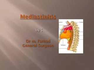
Mediastinitis by : Dr m. Farhad General Surgeon. ACUTE MEDIASTINITIS:. Typical Clinical Features of Acute Mediastinitis. Clinical Classification of Acute Mediastinitis. Etiologies and Clinical Settings. diagnosis. management. Complications of acute mediastinitis. ACUTE MEDIASTINITIS.

E N D
ACUTE MEDIASTINITIS: Typical Clinical Features of Acute Mediastinitis Clinical Classification of Acute Mediastinitis Etiologies and Clinical Settings diagnosis management Complications of acute mediastinitis
ACUTE MEDIASTINITIS Acute mediastinitis is rare and dramatic condition of a fulminating and usually fatal course Typical Clinical Features of Acute Mediastinitis . sudden and dramatic onset , with chills, high fever, and prostration. Patients are restless and irritable, Tachycardia, tachypnea, . severe substernal chest pain, worsened by breathing or coughing, and unrelieved by opiates. The pain may be referred into the neck and ear if the process involves the superior mediastinum , whereas posterior or inferior mediastinal involvement may cause radicular pain radiating around the chest and pain between the scapulae.
Signs: supraclavicular fullness and tenderness over the sternum or sternoclavicular joints, crepitus and other signs of mediastinal and subcutaneous emphysema may be prominent. Hamman's sign (a crunching sound synchronous with cardiac systole, heard over the anterior thorax) is characteristic but not always present. Later, tracheal deviation, jugular venous distention, and other signs of compression of mediastinal structures may appear.
Clinical Classification of Acute Mediastinitis Involvement of different mediastinal regions tends to have typical causes: infection in the superior mediastinum is most often the result of direct extension from neck infection; anterior mediastinal infection is typical after surgery or penetrating wounds to the anterior thorax; and posterior mediastinal abscesses are characteristic for tuberculous or pyogenic spinal infections.
1 : Mediastinitis Resulting from Visceral Perforation :Boerhaave's syndrome refers to esophageal rupture associated with forceful vomiting, classically after overeating or excessive drinking. It is the most familiar example of acute mediastinitis, clinical manifestations: In addition to the clinical manifestations described previously, hematemesis may be present before the actual rupture, and tends to diminish or stop after rupture occurs. Unilateral or bilateral hydropneumothorax is common and quickly progresses to empyema
The diagnosis of esophageal perforation ☻depends on an appropriate degree of clinical suspicion. ☻On the chest roentgenogram, ● the hallmarks are diffuse mediastinal widening ●presence of air in the mediastinum and elsewhere in soft tissues. ● Mediastinal air-fluid levels may be seen, ●pneumothorax or hydropneumothorax may be present. ☻CT can delineate these abnormalities more clearly.
ESOFAGOGRAMA • Opacificacióndel árbol bronquial de lóbulos inferiores, por paso del material de contraste al árbol bronquial. • Ensanchamiento mediastínico • Neumomediastino • Enfisema subcutáneo cervical
TC. SxBoerhaave • Aire en mediastino posterior (flecha) • Engrosamiento de la pared esofágica • Derrame pleural bilateral
The diagnosis is usually established by contrast studies, endoscopic examination, although percutaneous mediastinal aspiration, using a subxiphoid approach, is advocated by some as a means of earlier diagnosis ☻Successful management of frank, uncontained esophageal perforation : ●early surgical repair, drainage of the mediastinum and often the pleural space, ●administration of appropriate antibiotics, ●Percutaneous catheter aspiration of mediastinal abscesses, under CT guidance, IF infection is localized and the clinical setting is less urgent
☻Complications of acute mediastinitis after :esophageal rupture ● localized abscess formation, ● extensive pleural empyema, ● and persistent esophagocutaneous fistulas. ● Mortality reported due to acute mediastinitis after esophageal rupture has ranged from 10% to 20%to as high as 40% to 50% □Timing of surgical drainage has been of prime importance in determining the clinical outcome
☻Other potential iatrogenic causes of mediastinitis include: ● bronchoscopic perforation and migration of indwelling central venous catheters. ●use of laser and mechanical endobronchial procedures, in the setting of malignancy with chronic airway colonization or postobstructive pneumonia, add to the likelihood of potential mediastinal complications. ●Intravascular catheters may be another source of acute mediastinitis when the catheter tip erodes through the vessel wall into the mediastinum. Instillation of hyperosmotic,vesicant, or vasoactive substances via these catheters may induce a chemical, rather than an infectious, inflammation
Direct Extension of Infection from Other Sites: secondary to : oropharyngeal infection Infection originating in periodontal tissues in the tonsillar region, or after pharyngeal perforation extend via the prevertebral, visceral, or pretracheal spaces or in the carotid sheaths although the usual route is via the retropharyngeal space to the posterior mediastinum, • also named descending necrotizing mediastinitis, is perhaps the most clinically devastating form of the disorder. • Odontogenic infection is consistently the most common source of descending necrotizing mediastinitis
clinical signs : described before Radiological signs : widening of the retropharyngeal space, with or without associated air-fluid levels, anterior displacement of the tracheal air column, and loss of the normal cervical spine lordosis on lateral films of the neck. infections are mixed with both aerobic and anaerobic organisms
La principal infección es la angina de Ludwig • infección del segundo o tercer molar inferior que involucra a los espacios sublingual y submaxilar. • Puede diseminarse por el espacio faríngeo lateral >>> al espacio retrofaríngeo o vaina carotídea >>>> el mediastino.
Routine serial postoperative cervicothoracic CT imaging and aggressive reexploration and drainage guided by these imaging findings appear to reduce the mortality of this condition Although thoracoscopic and other percutaneous drainage procedures have been described and may be appropriate in selected patients, thorough open drainage and irrigation remain the standard approach. Treatment of descending necrotizing mediastinitis requires aggressive surgical drainage, usually via a cervical approach. thoracic exploration be reserved for cases in which the infection extends below the level of the fourth vertebral body or the tracheal bifurcation.
Rare cause of acute mediastinitis: with eroding neoplasms. Extension from anterior chest wall and neck infections has been described in injection drug users and acute purulent mediastinitis has also been reported after closed-chest cardiopulmonary resuscitation as a complication of vertebral or costal tuberculous Both gastric and esophageal ulcers have been reported as causes of mediastinitis, sometimes eroding directly into the pericardium.
Mediastinitis after Cardiac Surgery bacterial mediastinitis after median sternotomy for coronary artery bypass, valve replacement, correction of congenital heart disease after heart and heart-lung transplantation. as a complication of endoscopy,
risk factors Preoperative risk factors include advanced age and male gender, diabetes mellitus, obesity, the need for immunosuppressive therapy, smoking, obstructive lung disease, a history of previous sternotomy or mediastinal irradiation, and poorer preoperative cardiac dysfunction Perioperative risk factors include shaving rather than clipping for hair removal, the use of bilateral internal mammary artery grafts, a longer duration of the surgical procedure and of perfusion time, greater use of cautery or bone wax,
Postoperative risk factorsa low cardiac output state in the early postoperative period, and greater amounts of postoperative bleeding. accompanies coronary artery bypass, or if the patient requires more than 48 hours of mechanical ventilation postoperatively The pathogenesis of mediastinitis following sternotomy is debated, although most cases appear to result from direct contamination of the mediastinum at the time of operation.
In the acute stages, the mediastinal structures are involved with pliable fibrinous exudates, and osteomyelitis, if present, is confined to the wound margins. Subacute infections are characterized by increasingly dense adhesions entrapping the visceral organs, sinus tract formation, and more extensive sternal bone involvement.
Prevention: infection control and careful asepsis in the operating room remain the most effective means of prevention prophylactic antibiotics are widely used in the perioperative management of cardiac surgery patients. The prophylactic intranasal application of mupirocin ointment has been shown to reduce by 50% the rate of Staphylococcus aureus nosocomial infections
Mediastinitis may occur as early as 3 days or as long as 6 months after surgery although most cases occur within 2 weeks. The bacteriology of postoperative mediastinitis In early prosthetic valve endocarditis. Staphylococcus epidermidis and S. aureus have been the most frequent organisms Anaerobes and gram-negative bacilli are rare, Candida species and atypical mycobacteria (especially Mycobacterium chelonae and Mycobacterium fortuitum) are infrequently reported. Infection with the last two groups tends to be more indolent
the clinical course consists of fever and systemic signs, followed by bacteremia and local signs of wound infection Thediagnosis is usually made at the time of reexploration of the sternotomy wound and rests on a heightened clinical suspicion in the appropriate setting. diagnostic tests gallium scanning, CT, and ultrasonography. CT is particularly helpful in identifying and discerning soft-tissue swelling, fluid collections, and sternal erosion or dehiscence.
Patients with fever, positive blood cultures, and wound abnormalities in the post-sternotomy period should probably be explored. therapy for post-sternotomy mediastinitis consists of early surgical exploration, débridement and drainage, irrigation, and prolonged administration of systemic antibiotics.
"Primary Mediastinitis": Inhalational Anthrax Anthrax, caused by infection with Bacillus anthracis, is primarily a disease of cattle, sheep, and goats and is most prevalent in the Middle East, although it is now recognized as an important disease of bioterrorism inhalational anthrax, or woolsorter's disease, is contracted by inhaling B. anthracis spores from animal sources. Inhalation of anthrax spores into the distal air spaces is followed by ingestion by alveolar macrophages and transport to the mediastinal lymph nodes. A hemorrhagic mediastinitis rapidly evolves, followed by bacteremia, overwhelming sepsis, and usually death.
Clinically patients typically experience a biphasic illness with an initial insidious flulike illness lasting 2 to 4 days and characterized by fever, malaise, myalgia, and nonproductive cough. This is followed by afulminant phase of acute mediastinitis, with respiratory distress, chest pain, cyanosis, and prostration. The chest radiograph and CT scan typically show mediastinal widening and pleural effusions
The diagnosis is established by demonstration of gram-positive, boxcar-shaped bacilli in tissue or body fluid specimens or in the blood buffy coat A direct fluorescent antibody test, polymerase chain reaction, and serologic tests are available for confirmation.
High-dose intravenous penicillin has traditionally been the treatment of choice,although penicillin-resistant strains have been reported ,Cephalosporin resistance is typical, and most patients in the bioterrorism outbreak were treated with multiple agents, including a fluoroquinolone. Inhalational anthrax has historically been a devastating disease even with appropriate treatment in the bioterrorism outbreak, prompt diagnosis and initiation of antibiotic therapy plus aggressive drainage of mediastinal and pleural collections resulted in survival of 6 of the 10 patients.