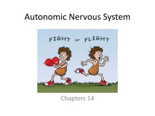
Autonomic Nervous System
700 likes | 729 Views
Learn about the autonomic nervous system controlling visceral functions like heart, GIT, and bladder, through sympathetic and parasympathetic pathways. Discover how higher centers in the brain influence autonomic control.

Autonomic Nervous System
E N D
Presentation Transcript
Autonomic Nervous System Introduction Chapter 61,pg773,Guyton 13th edition
Autonomic Nervous System • Visceral functions • Heart • GIT • Urinary Bladder • Rapidity & Intensity
Autonomic Nervous System • Higher control: • Spinal cord • Brain Stem • Hypothalamus • portions of the cerebral cortex,esp. limbic cortex, can transmit signals to the lower centers & can influence autonomic control.
ANS also operates through visceral reflexes i.e. subconscious sensory signals from visceral organs autonomic ganglia, the brain stem, or the hypothalamus return subconscious reflex responses directly back to the visceral organs to control their activities.
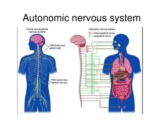
Efferent autonomic signals are transmitted to the various organs of the body through two major subdivisions • Sympathetic nervous system • Parasympathetic nervous system
In the central nervous system, a collection of neuron cell bodies is called a nucleus. In the peripheral nervous system, a collection of neuron cell bodies is called a ganglion.
Sympathetic system is also referred to • fear flight n fight response. • It extends between T1-L2, also called thoraco-lumber division.
Physiological Anatomy of the Sympathetic Nervous System • (1) one of the two paravertebral sympathetic chains of ganglia that are interconnected with the spinal nerves on the side of the vertebral column. • (2) prevertebral ganglia(the celiac, superior mesenteric,aortico-renal, inferior mesenteric,and hypogastric). • (3) nerves extending from the ganglia to the different internal organs
Each sympathetic pathway from the cord to the stimulated tissue is composed of two neurons, • preganglionic neuron • postganglionic neuron • in contrast to only a single neuron in the skeletal motor pathway • The cell body of each preganglionic neuron lies in the intermediolateral horn of the spinal cord; its fiber passes through a ventral root of the cord into the corre-sponding spinal nerve.
After the spinal nerve leaves the spinal canal, the preganglionic sympathetic fibers leave the spinal nerve and pass through a white ramus into one of the ganglia of the sympathetic chain.
The fibers then can take one of the following three courses: • (1) they can synapse with postganglionic sympathetic neurons in the ganglion that they enter; • (2) they can pass upward or downward in the chain and synapse in one of the other ganglia of the chain; or
(3) they can pass for variable distances through the chain and then through one of the sympathetic nerves radiating outward from the chain, finally synapsing in a peripheral sympathetic ganglion.
Sympathetic Nerve Fibers in the Skeletal Nerve • Some of the postganglionic fibers pass back from the sympathetic chain into the spinal nerves through gray ramiat all levels of the cord. • These sympathetic fibers are all very small type C fibers, and they extend to all parts of the body by way of the skeletal nerves.
They control the blood vessels, sweat glands, and piloerector muscles of the hairs. • About 8 percent of the fibers in the average skeletal nerve are sympathetic fibers.
Segmental Distribution of the Sympathetic Nerve Fibers. • The sympathetic pathways that originate in the dif-ferent segments of the spinal cord are not necessarily dis-tributed to the same part of the body as the somatic spinal • nerve fibers from the same segments. Instead, the
Sympathetic fibers from cord segment • (1) T1 generally pass up the sympathetic chain to terminate in the head; • (2) from T2 to terminate in the neck; • (3) from T3, T4, T5, and T6 into the thorax; • (4) from T7, T8, T9, T10, and T11 into the abdomen; • (5) from T12, L1, and L2 into the legs. • This distribution is only approximate and overlaps greatly.
The distribution of sympathetic nerves to each organ is determined partly by the locus in the embryo from which the organ originated. • For instance, the heart receives many sympathetic nerve fibers from the neck portion of the sym-pathetic chain because the heart originated in the neck of the embryo before translocating into the thorax.
Special Nature of the Sympathetic Nerve Endings in the Adrenal Medullae. • Preganglionic sympathetic nerve fibers pass, without synapsing,all the way from the intermediolateral horn cells of the spinal cord, through the sympathetic chains, then through the splanchnic nerves, and finally into the two adrenal medullae.
There they end directly on modified neuronal cells that secrete epinephrine and norepinephrine into the blood stream
Physiological Anatomy of the Parasympathetic Nervous System • Parasympathetic fibers leave the central nervous system through cranial nerves III, VII, IX, and X. • Additional parasympathetic fibers leave the lowermost part of the spinal cord through the 2nd and 3rd sacral spinal nerves and occasionally the S1 and S4 nerves.
About 75 percent of all parasympa-thetic nerve fibers are in the vagus nerves(cranial nerve X), passing to the entire thoracic and abdominal regions of the body.
Parasympathetic fibers in the III cranial nerve go to the pupillary sphincter and ciliary muscle of the eye. • Fibers from VII cranial nerve pass to the lacrimal, nasal, and submandibular glands, and • Fibers from the IX cranial nerve go to the parotid gland.
The sacral parasympathetic fibers are in the pelvic nerves,which pass through the spinal nerve sacral plexus on each side of the cord at the S2 and S3 levels. • These fibers then distribute to the descending colon, rectum, urinary bladder, and lower portions of the ureters.
Basic Characteristics Cholinergic & Adrenergic Fibers
Sympathetic Parasympathetic Cholinergic Cholinergic Cholinergic Sweat Glands, Piloerecror Muscle, A few Blood Vessels Cholinergic Adrenergic
Acetylcholine Synthesis From Choline In terminal nerve endings Destruction Acetylcholinesterase Norepinephrine Synthesis From Tyrosine In axoplasm of nerve endings Removal Reuptake Diffusion into body fluid n into Blood Enzymes MAO(in nerve endings) catechol-O-methyl transferase(in tissues) Metabolism of Neurotransmitters
Norepinephrine and epinephrine secreted into the blood by the adrenal medullae remain active until they diffuse into some tissue, where they can be destroyed by catechol-O-methyl transferase; this action occurs mainly in the liver.
Circulation of neurotransmitters • Acetylcholine: • Local • Noradrenalin/ Norepinephrine: • Local • Blood • Adrenalin/ Epinephrine • Blood • Hormone
Other Neurotransmitters • Dopamine • GnRH (Gonadotrophin Releasing Hormone) • Co-transmitters • VIP • ATP • Neuropeptide Y