Phineas Gage: Case Study on Brain Damage and Morality Change
360 likes | 461 Views
Explore Phineas Gage's famous case of brain damage impacting his personality and cognitive functions. Understand how brain imaging techniques like CT scans and PET scans reveal brain function and metabolism. Learn about glucose utilization by the brain and the impact of brain injuries on behavior.

Phineas Gage: Case Study on Brain Damage and Morality Change
E N D
Presentation Transcript
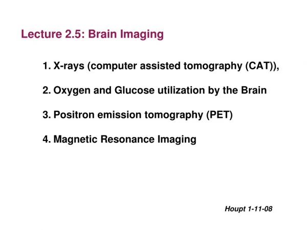
Lecture 2.5: Brain Imaging X-rays (computer assisted tomography (CAT)), Oxygen and Glucose utilization by the Brain Positron emission tomography (PET) Magnetic Resonance Imaging Houpt 1-11-08
The most famous case of brain damage that causes a change in morality remains that of Phineas Gage, a railroad laborer in Vermont who, one day in the fall of 1848, suffered a horrific on-the-job injury. Gage, the foreman of a crew laying track outside the town of Cavendish, was tamping black powder into a hole drilled in rock when he apparently struck a spark. In a flash of explosion, the tamping iron, a three-and-a-half-foot-long bar an inch in diameter, blew through his left cheek and clean out the top of his head, landing some 30 yards behind him. "It essentially severed the front third of his brain," Eslinger says. "The surgeon who came to the scene described that he insert a could finger through either side of the wound and actually touch them. It was just this clean hole." www.rps.psu.edu/ indepth/brainscans1.htm
Phineas Gage's skull and life mask Credit Warren Anatomical Museum, Francis A. Countway Library of Medicine
X-rays (computer assisted tomography (CAT) Reconstruction of the lesion incurred by Phineas Gage, in which an iron bar was driven through his prefrontal cortex as a result of a blasting accident.
Amazingly, Gage survived, and was in fact strong enough to resume work in less than a year. His basic mental faculties—motor skills, memory, speech—were essentially intact. What had changed, profoundly and irrevocably, was his personality. Where before the accident, Gage had been regarded as an excellent foreman, thoughtful, shrewd with money, and well-spoken, afterward he was described as "fitful, irreverent, and grossly profane," and acting with little regard for others. His friends said he was "no longer Gage." www.rps.psu.edu/ indepth/brainscans1.htm
Methods for analyzing brain function: spatial and temporal resolution
Two ways to detect change in brain activity (without using electrodes): • 1. Change in blood flow: • more active brain regions have increased blood flow • 2. Change in glucose utilization: • more active regions use more glucose • (note: both these approaches are relative changes, measured against some baseline)
Glucose is energy source for the brain Brain = 2% of body weight Brain uses 25% of glucose & 20% of oxygen glucose and oxygen used to generate ATP to drive most cellular processes Can track increased neural activity by increased glucose and oxygen utilization.
Can’t label plain glucose, just get radioactive CO2 So accumulated 2DG means cell (trying) to use glucose 2-Deoxy-Glucose (2DG) can track metabolic activity… label with radioactive 13C
To detect metabolism with 2DG: post mortem radioactive 13C-2DG is administered, the brain is removed, and tissue sections are apposed to film; an autoradiogram is created when the radiation exposes the film. in vivo radioactive fluro-2DG is administered intravenously. 2DG sequested in metabolically active regions is detected with Positron Emission Tomography (PET scan) to make 3D images of the brain.
PET Scan (in vivo example) accumulation of radioactive 2DG in living person
Autoradiogram example (Postmortem) 1. Inject monkey with 13C-2DG 2. Project pattern onto screen in front of monkey 3. Let monkey watch screen for 1 hour 4. Remove brain and expose visual cortex to film So, are these neurons that respond to visual input?
Blood brain barrier prevents diffusion of glucose into brain General Capillary Brain Capillary
How does Glucose get to neurons? Blood-brain barrier (BBB) prevents diffusion of large, polar molecules like glucose from the blood to the extracellular space of the brain. There are glucose transport proteins (GLUTs) in the endothelial cells that move glucose by facilitated transport across the BBB. The astrocytes have a large number of GLUTs to take up glucose. Neurons have a specific GLUT-3 to take up some glucose. Astrocytes metabolize glucose to lactate, which is released and taken up by neurons. (Neurons and astrocytes in the feeding centers of the hypothalamus have GLUT-2 transporters, like the pancreas, to monitor glucose levels).
Glucose Transporters Glucose Lactate
Test in a system with clean separation of glia and neurons: photoreceptor neuron glial cells photo Honey bee photoreceptor neurons are arrayed in hexagonal “rosettes”, surrounded by glial cells.
photoreceptor neuron glial cells autorad After incubation with 2DG, the neurons accumulate almost no glucose, but surrounding glia cells are heavy users of glucose.
Glial cells partially metabolize glucose -> lactate -> neurons
Release of K+, adenosine, lactate and nitric oxide by neurons and glia causes local vasodilation and increase in oxygenated hemoglobin
PET scan Different isotopes can be used for different measures: e.g, 18F-deoxy-Glucose for metabolism, or 15O-water for blood flow
(Flashing light) Visual cortex at back of brain
Synopsis of MRI Put subject in big magnetic field Transmit radio waves into subject (2-10 ms) Turn off radio wave transmitter Receive radio waves re-transmitted by subject Convert measured RF data to image James Voyvodic, Ph.D
Hydrogen atoms are best for MRI • Biological tissues are predominantly 12C, 16O, 1H, and 14N • Hydrogen atom is the only major species that is MR sensitive • Hydrogen is the most abundant atom in the body • The majority of hydrogen is in water (H2O) • Essentially all MRI is hydrogen (proton) imaging James Voyvodic, Ph.D
Two types of MRI image T1 image: H atoms in non-aqueous environment T2 image: H atoms surrounded by water Bone, white matter, fat looks bright Fluid spaces, gray matter looks bright
Increased blood oxygenation level detection (BOLD) by MRI oxygenated hemoglobin can be detected by MRI. so increased oxyhemoglobin reflects increased blood flow which reflects increased brain activity. Response to light
http://science.sciencemag.org/content/353/6303/1030.full https://www.youtube.com/watch?time_continue=131&v=N9QQxa6eLPc
D) Dog auditory regions responsive to speech (table S2). The color bar shows the range for one-sample t-test scores (12 degrees of freedom) for the speech (Pp + Pn + Np + Nn) > silence contrast.
Praise Words - Positive Intonation Praise Words - Neutral Intonation Neutral Words - Positive Intonation Neurtal Words - Neutral Intonation Reward Pathway Praise Words - Neutral Intonation Dopamine


