Download
1 / 45
460 likes | 1k Views
Breast Lumps. Presented by :- Divya Divakaran Foundation Year 2 Doctor. Contents. Anatomy of Breast History taking Clinical examination Specific considerations Triple assessment Diagnosis Management What about men?. Anatomy of Breast. Introduction to breast :

E N D
Breast Lumps Presented by :- Divya Divakaran Foundation Year 2 Doctor
Contents • Anatomy of Breast • History taking • Clinical examination • Specific considerations • Triple assessment • Diagnosis • Management • What about men?
Anatomy of Breast • Introduction to breast : • Breasts (mammary glands) = modified sweat glands
The breast is composed of glandular, ductal, connective, and adipose tissue. The mammary glands are modified sweat gland and are composed of 15-20 lobules, each drained by a lactiferous duct. Each lactiferous duct independently drains on the nipple. Areola surrounds nipple • In men, little fat is present in the breast, and the glandular system normally does not develop.
Lie in superficial fascia anterior to deep fascia of pec. major BOUNDARIES :- • Bounded by the clavicle superiorly • Infra-mammary fold inferiorly • The sternum medially • Lateral border of the latissimus muscle laterally
Coopers ligament • The glands are firmly attached to the skin by connective tissue structures known as Cooper's ligaments or suspensory ligaments. Coopers ligament help maintain the structural integrity. They are named for Astley Cooper, who first described them in 1840. It is these ligaments which pull on the skin, creating the characterisctic dimpling (or peau d'orange) associated with malignancy
Four Quadrants of the Breast • Upper outer (superolateral) quadrant • Upper inner (superomedial) quadrant • Lower outer (inferolateral) quadrant • Lower inner (inferomedial) quadrant
The French term peau d'orange means skin of an orange • Advaned malignancy leads to infiltration and shortening of Cooper’s ligament • Leads to irregular dimpling of skin or retraction of nipple
Arteries : Predominatly Internal mammary, lateral thoracic, thoracoacromial, posterior intercostal • Veins : Mainly Axillary (subclavian, intercostal, internal thoracic) • Lymphatics : Axillary, parasternal, inferior phrenic nodes • Nerves : 4th-6th intercostal nerves
The lymphatic drainage of the breast deserves special attention, due to its role in the metastasis of cancer cells. The majority of lymph (>75%), particularly from the lateral quadrants, drains to the axillary lymph nodes. The remainder of lymph drains to either the parasternal nodes or the opposite breast
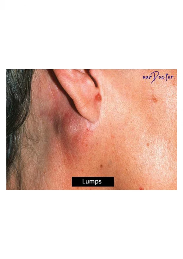
History taking : The Lump • Onset : when was the lump first noticed • Location : which side - right or left • Single or multiple : how many ? • Unilateral or bilateral • Duration : since when did the pt notice the lump • Progression : Has it changed in size (ca) • Is there any pain : type, severity (painless in ca ) • Association with menstrual cycle
Skin changes, nipple discharge or retraction • Axillary / supraclavicular swelling • Previous breast cancer • H/o trauma, SOB, bone pain, fever or weight loss
Risk factors • Female sex, older age • Family history of breast ca • Oral contraceptive pill / HRT • Cycles (early menarche or late menopause) • Pregnancy : lack of child bearing • Lack of breast feeding • Smoking, alcohol intake • High fat diet
Past medical history • Surgical history • Drug history • Allergies • Social history : support, activity level, smoking, alcohol, drugs
Examination • Specific considerations : • Chaperone must be present • Explain to them what the examination will entail and gain the patients consent
Examination • Inspection • Palpation • Auscultation
Inspection • Inspect the patient in upright position • Make a general inspection of both breasts. Look for any asymmetry, scars, obvious lumps or nipple abnormalities (e.g. inversion or discharge) • You should also comment on any skin changes (peaud’orange, eczema).
Ask the patient to place her hands above her head and repeat the inspection • Look for any obvious mass
Palpation • Start on the “normal” side first • Ask the patient to place her hand behind her head on the side you are examining • Systematically examine all areas of the breast with your hand laid flat on the breast. Start from outside and work towards the nipple. Imagine that the breast is a clock face and examine at each ‘hour’
LUMP : size, shape, position,consistency, surface,overlying skin • Don’t forget that the breast tissue extends towards the axilla in the ‘axillary tail’ • Ensure you ask the patient if she experiences any pain during examination
Examine the other breast in the same manner • Ask the patient to squeeze both nipples
Lymph node examination • Examine both axillae for any enlarged lymph nodes • Whilst examining the patient’s axilla, you should fully support the weight of that arm with yours • Examine the axilla with your other hand
Ensuring that you feel all four walls (anterior, posterior, medial and lateral) as well as feeling into the apex of the axilla • Repeat this on the other side • Palpate the supraclavicular fossa on both sides to check for lymphadenopathy • Finalyauscultate the chest
Triple assessment • Clinical findings • Radiological findings (Mammography/ USS) • Biopsy : Histology/cytology • If there is any abnormality detected in the examination, or imaging, then biopsies are taken. This can be in the form of FNAC (Fine Needle Aspiration Cytology) or a core (Tru-Cut) biopsy.
Breast Cancer • Commonest Cancer among women in UK. About 48,000 women get breast cancer in Britain each year. • Breast cancer originates from breast tissue, most commonly from the inner lining of milk ducts (ductal ca )or the lobules that supply the ducts with milk (lobular ca) • Ductal Ca 90%, lobular Ca 10%
Increase in incidence with age • Associated with mutations in the breast cancer susceptibility genes BRCA1 or BRCA2 • Risk factors : Smoking, alcohol, OCPs, HRT • C/F : painless lump, discharge, skin changes
Stages of breast ca Common symptoms for breast cancer in both men and women are: • Swelling or redness in the skin on or around the breast area • A change in size or shape of one or both of the breast • A lump or mass in the breast or near the under arm • Changes in the appearance of nipple • Discharge of fluid other than milk out of the nipple
Types of breast Cancer • Breast cancer is often divided into non-invasive and invasive types • Non-invasive breast cancer is also known as cancer or carcinoma in situ. This cancer is found in the ducts of the breast and has not developed the ability to spread outside the breast. This form of cancer rarely shows as a lump in the breast and is usually found on a mammogram
Invasive cancer has the ability to spread outside the breast • Invasive ductal breast cancer accounts for about 80% of all cases of breast cancer
Spread It is possible for breast cancer to spread to other parts of the body, usually through the lymph nodes or the bloodstream. If this happens, it is known metastatic breast cancer. It metastasis to the lymph nodes, lungs, liver, bones etc
Treatment : Breast cancer is treated using a combination of surgery (lumpectomy or mastectomy), chemotherapy and radiotherapy • There is a good chance of recovery if it is detected in its early stages.
Fibroadenoma • Fibroadenoma of the breast is a benign tumor composed of two elements : epithelium and stroma • Fibroadenomas are called breast mouse owing to their high mobility in the breast • Painless, firm and mobile • In young women of child bearing year
Hormone-dependent and frequently regress after menopause • Investigation : needle biopsy • Treatment : surgical excison
Benign breast conditions • Mastalgia : cyclical and non cyclical • Cyclical mastalgia : painful nodularity associated with ovulation, hyperplasia secondary to E2 • Non cyclical mastalgia : trauma, mastitis, shingles, diurectics
Mastitis • Inflammation of breast tissue • Staph aureusis the main organism • 2-10% in lactating women at 2-4 weeks post partum • Treated with antibiotics • 90% cured, 10% abscess formation (drainage)
Duct ectasia • Blockage of the lactiferous duct The duct widening is commonly believed to be a result of secretory stasis (stagnant colostrum) or subject to hormonal interactions or non specific
pre-menopausal age • Mimic breast cancer, noncyclic breast pain • Clinical features : pain, nipple retraction or nipple discharge • Self limiting and not indicated for surgery
Pappiloma • Benign lesion • Types: central and peripheral • Central type: single solitary lesion develops near nipple, seen nearing menopause Peripheral type : multiple papillomas in the periphery of breast seen in young women • C/F: Bloody nipple discharge, masses are too small to be palpated
Higher risk of malignant change • Investigation : galactogram +/- biopsy, not shown in mammograpghy due to small size • Excision is sometimes performed (benign)
Men !!! • Less than 1% • Peak incidence at 60yrs • Worst prognosis
Men • Gynaecomastia : steroids, hormonal therapy, spiranolactone, CCB, testicular tumours, pituitary tumours, obesity and in elderly
Summary • History • Think risk factors • Likely diagnosis • Practise examination