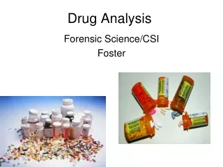
Drug Analysis
Drug Analysis. Drug Identification. Screening or presumptive tests Spot or color tests Microcrystalline test — a reagent is added, producing a crystalline precipitate that is unique for a certain drug Chromatography. Confirmatory tests Spectrophotometry Ultraviolet (UV) Visible

Drug Analysis
E N D
Presentation Transcript
Drug Identification Screening or presumptive tests Spot or color tests Microcrystalline test— a reagent is added, producing a crystalline precipitate that is unique for a certain drug Chromatography Confirmatory tests Spectrophotometry • Ultraviolet (UV) • Visible • Infrared (IR) Mass spectrometry
Drug Identification, continued Screening or presumptive tests only tell that the drug is possibly present. Confirmatory tests tell that the drug is positively present. (Screening tests are easier, cheaper, and quicker to use.)
Presumptive Color Tests Marquis—turns purple in the presence of most opium derivatives and orange-brown with amphetamines Dille-Koppanyi—turns violet-blue in the presence of barbiturates Duquenois-Levine—turns a purple color in the presence of marijuana Van Urk—turns a blue-purple in the presence of LSD Scott test—color test for cocaine; blue
Chromatography A technique for separating mixtures into their components Includes two phases—a mobile one that flows past a stationary one The mixture interacts with the stationary phase and separates
Types of Chromatography Paper Thin-layer (TLC) Gas (GC) Pyrolysis gas (PGC) Liquid (LC) High-performance liquid (HPLC) Column
Paper Chromatography Stationary phase—paper Mobile phase—a liquid solvent Capillary action moves the mobile phase through the stationary phase.
Thin-layer Chromatography Stationary phase—a thin layer of coating (usually alumina or silica) on a sheet of plastic or glass Mobile phase—a liquid solvent
Retention Factor (Rf) This is a number that represents how far a compound travels in a particular solvent. It is determined by measuring the distance the compound traveled and dividing it by the distance the solvent traveled. If the Rf value for an unknown compound is close to or the same as that for the known compound, the two compounds are likely similar or identical (a match).
Gas Chromatography Phases Stationary—a solid or a viscous liquid that lines a tube or column Mobile—an inert gas like nitrogen or helium Analysis Shows a peak that is proportional to the quantity of the substance present Uses retention time instead of Rffor the qualitative analysis
Uses of Gas Chromatography Not considered a confirmation of a controlled substance Used as a separation tool for mass spectroscopy (MS) and infrared spectroscopy (IR) Used to quantitatively measure the concentration of a sample. (In a courtroom, there is no real requirement to know the concentration of a substance. It does not affect guilt or innocence.)
Confirmatory Tests: Spectroscopy Spectroscopy—the interaction of electromagnetic radiation with matter Spectrophotometer—an instrument used to measure and record the absorption spectrum of a chemical substance
Spectrophotometry Components A radiation source A frequency selector A sample holder A detector to convert electromagnetic radiation into an electrical signal A recorder to produce a record of the signal Types Ultraviolet Visible Infrared
Infrared Spectrometry Material absorbs energy in the near-IR region of the electromagnetic spectrum Compares the IR light beam before and after it passes through a transparent sample Result—an absorption or transmittance spectrum Gives a unique view of the substance; like a fingerprint
Mass Spectrometry Gas chromatography has one major drawback: It does not give a specific identification. Mass spectrometry cannot separate mixtures. By combining the two (GC-MS), constituents of mixtures can be specifically identified.
Mass Spectrometry, continued In a mass spectrometer, an electron beam is directed at sample molecules in a vacuum chamber. The electrons break apart the sample molecules into many positive-charged fragments. These are sorted and collected according to their mass-to-charge ratio by an oscillating electric or magnetic field.
Mass Spectra Each molecular species has its own unique mass spectrum.
IR Spectrophotometry and Mass Spectrometry Both work well in identifying pure substances. Mixtures are difficult to identify in both techniques. Both are compared to a catalog of knowns.