Download
1 / 64
810 likes | 1.24k Views
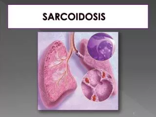
SARCOIDOSIS. SARCOIDOSIS. Definition : Idiopathic systemic disorder characterized by accumulation of lymphocytes and monocytes in many organs forming noncaseating, epitheloid granuloma and subsequent conformational changes in the involved organs Etiology : unknown

E N D
SARCOIDOSIS • Definition: Idiopathic systemic disorder characterized by accumulation of lymphocytes and monocytes in many organs forming noncaseating, epitheloid granuloma and subsequent conformational changes in the involved organs • Etiology: unknown • Extent of involvement : systemic • Clinical course : variable from asymptomaticdisease with spontaneous resolution to progressive disease with organ system failure • Symptoms: dependent on site of involvement
Epidemiology • Common in : N. Europe (especially Scandinavia, Ireland, Great Britain), N. America, and Japan • Low incidence: China, India, Africa, Russia • Peak age of incidence: 20’s and 30’s • Sex prevalence : women > men • In USA : blacks > whites • Worldwide: 80% of affected patients are white
Etiopathogenesis • Genetic susceptibility • Environmental factors • Triggers an immune response (Th type 1)
Genetic factors • Prevalence in certain race • Familial clustering • HLA -A1, -B8, and -DR3 • HLA B22 in Italians • HLA DR-17 good prognosis in Scandinavians; protracted course with DR 15 and 16 • DR5j Japanese patients have poor prognosis • Negative association: HLA B12 and -DR4
Environmental agents • 1969 Mitchell and Reese - ? Infectious agent • Non-infectious agents ? Aluminum, berrylium etc. • Mycobacterial ?
Granulomatous reaction • T cells predominantly CD4 accumulate • Releases IFN-gamma, IL-2 and other cytokines • Macrophages are recruited and release its own inflammatory mediators (TNF, IL12, IL15, growth factors) • CD45RO + Th1 type lymphocyte is activated • Granuloma formation
Signs and Symptoms • Depend on the site • Fever, fatigue, weight loss, arthralgias (1/3) • Persistent fever seen in liver involvement • Peripheral lymphadenopathy usually asymptomatic • Cough and dyspnea less often seen
SARCOIDOSISSystemic Involvement • Lung lesions – 95% • Thoracic lymph nodes – 50% • Skin lesions – 30% • Eyes – 30%
SARCOIDOSISSystemic signs • Hilar adenopathy on chest x-ray • Lung infiltrate • Erythema nodosum • Arthritis
Lung involvement • In 90% - 95%of cases • Dyspnea, dry cough, and chest pain (1/3) • Primarily involves the parenchyma • Lymph node involvement, and airway lesions (larynx, trachea and bronchi) may also be involved; 20% asthma-like • Mediastinal adenopathy on routine x-ray • Bilateral hilar and right paratracheal adenopathy universally seen
Lung involvement • Pulmonary infiltrate may have a diffuse, fine, ground-glass appearance • Fibrosis, cystic changes, and cor pulmonale in late progressions • Uncommon manifestations include pleural effusion, pleural thickening, pneumothorax • cavity formation, lymph node (LN) calcification
Radiographic stages • Stage 0, no intrathoracic finding • Stage 1 • Bilateral hilar adenopathy, often accompanied by paratracheal node enlargement • 80% has regression of hilar nodes in 1-3 years • Stage 2 • Bilateral hilar adenopathy and interstitial infiltrates (upper lung zone more than lower) • Mild to mod symptoms • can undergo spontaneous resolution • Stage 3 • Interstitial disease with shrinking hilar nodes, upper lung zone interstitial opacities • Stage 4 • Advanced fibrosis
CXR Findings • Stage 1- Bilateral hilar adenopathy (80% resolution) • Stage 2- Hilar adenopathy + parenchymal infiltrates (50% resolution) • Stage 3- Parenchymal infiltrates (30% resolution) • Stage 4- Advanced fibrosis Sarcoidosis stage 1
CT Scan • Mediastinal and hilar adenopathy • Mid to upper lung predominance • Nodules along brochi, vessels or subpleural • Consolidation or ground glass opacity • Fibrosis with distortion of lung architecture
Stage I Stage II
Stage III Stage IV
Studies to evaluate pulmonary sarcoidosis • Imaging study with CXR, CT • Lung function tests – restrictive pattern, reduction in DLCO, endobronchial sarcoidosis presents obstructive pattern • Radiotracer scanning – staging the alveolitis in interstitial lung disease, unclear role • Broncho-alveolar lavage (BAL) – adjunctive measure to support the diagnosis. CD4:CD8 ratio • These support the diagnosis but not confirmatory • Differ.diagnosis of pulmonary involvement – hypersensitivity pneumonitis, eosinophilic granuloma, collagen vascular disease, pneumoconiosis, chronic beryllium lung dz, infections
Lung Function Tests • Lung function tests show restriction, decreased compliance, and impaired diffusing capacity • Co2 retention is uncomon, but airway obstruction is common in endobronchial disease and late states with pulm. fibrosis • Serial spyrometries are important for guiding treatment
Perpheral Lymph NodesInvolvement • Most common : cervical, epitrochlear, axillary, and inguinal nodes • Seen in 1/3 of patients • discrete, movable and non-tender • Do not ulcerate and form draining sinuses
Myocardial involvement • Myocardial involvement 5-10% • arrhythmias, heart failure (restrictive type), conduction abnormalities • The risk of cardiac dysfunction or sudden death in these patients is low (those with positive thallium-201 imaging) • endomyocardial biopsy confirms the diagnosis • need to exclude coronary artery disease
Eye involvement • In 15-25% of cases : • Anterior uveitis - the most common form of ocular sarcoidosis • - congestion, photophobia and ocular • discomfort • Heerfordt’s syndrome or uveoparotid fever - anterior uveitis + • parotitis, fever and facial palsy • Posterior uveitis - vitreous infiltrates, choroidal nodules, • periphlebitis, retinal hemorrhage, and • papilledema • Conjunctivitis - superficial congestion
Ocular Involvement • Anterior segment lesions (30%) • Conjunctival granuloma • Lacrimal gland involvement/dry eye • Acute or chronic uveitis • lesions described as ‘mutton fat’ because they are large and greasy
Ocular Involvement • Posterior segment lesions (20%) • Patchy venous sheathing • Cellular infiltrate around vessels • Chorioretinal granulonmas • Vasculitis including occlusive causing:- • Neovascularisation • Infiltrate in vitreous (vitritis) including cell clumps (snowballs)
Ocular Involvement Systemic steroids may be necessary in patients with posterior segment disease where vision is threatened, especially if optic nerve is involved
Skin disease • In chronic sarcoidosis 15-20% • plaques, papules, subcutaneous nodules • keloid formation in atrophic scars • Nasal and conjunctival mucosal granulomas may occur • erythema nodosum (EN) with fever and arthralgias seen often in Europeans; • EN + bilateral hilar lymphadenopathy =Lofgren’s syndrome, portends a good prognosis
Skin disease Lupus pernio violaceous, chronic and disfiguring lesions of the ears, nose and cheeks Lupus pernio affecting the nose – a chronic progressive cutaneous sarcoid
Neurologic Disease • In 5-10% of cases: • Unilateral facial nerve palsy - most common • Almost any structure can be involved • Hypotalamus-pituitary axis involvement can cause hyperprolactinemia and diabetes insipidus.
SARCOIDOSISSystemic signs • Facial palsy • Salivary gland enlargement
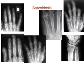
Joint involvement • Acute polyarthritis may be prominent • Chronic periarticular swelling and tenderness due to osseous changes in phalanges
Liver and other organs • Hepatic granulomas in biopsy in 50-80% of patients with normal liver function • Hepatomegaly in < 10% • Severe liver disease and jaundice are rare manifestations •Myopathy, splenomegaly, lacrimal gland, parotid gland, bone involvement
SARCOIDOSISInvestigations • Leukopenia frequent • Serum uric acid high, but gout is rare • Alkaline phosphatasis and GGT may be high if liver involved • Hypercalcemia +/- hypercalciuria due to calcitriol from macrophages • Depression of delayed hypersensitivity is characteristic: negative (or false neg) tuberculin skin test
SARCOIDOSISInvestigations • Serum angiotensin-converting enzyme (ACE) – elevated in active sarcoidosis • Mantoux test – caution in patients who have had BCG vaccination. Test may be negative. • Lung function tests - restriction
Broncho-alveolar lavage(BAL)Gallium scanning • CD4/CD8 ratio is elevated in BAL in sarcoidosis but reduced in hypersensitivity pneumonitis • whole-body gallium scanning is sensitive, but not specific • Symetric uptake in mediastinal and hilar nodes (lambda sign) and in lacrimal, parotid and salivary glands (panda sign) • Pathognomonic for sarcoidosis
SARCOIDOSISInvestigations Gallium scan showing increased uptake in the lacrimal and parotid glands and pulmonary regions in a patient with active sarcoidosis
Serum ACE • Serum ACE activity elevated in 40- 90% due to macrophage activity, but nonspecific since hystoplasmosis, acute milliary TB, hepatitis, and lymphomas also have this finding (5% false +) • Lacks diagnostic specificity and poor prognostic value in identifying patients with progressive disease • Tissue ACE activity is highest in sarcoid lymph nodes rather than in pulmonary tissues
Kveim-Siltzbach test • Rarely used in practice • Intradermal injection of homogonized tissue of organs involved with sarcoidosis causes delayed cutaneous reaction in 4-6 weeks • Within granulomas are multi-nucleated giant cells called with stellate inclusions called asteroid bodies and laminated calcificcations called Schaumann’s bodies
Questions • Do we need a biopsy to diagnose sarcoidosis? Where to biopsy? • What markers are available to follow disease progression of sarcoidosis? • What medications other than steroids are available for treatment?
Biopsy • Confirmation of diagnosis • Palpable lymph nodes • Subcutaneous nodule • Cutaneous lesion • Enlarged parotid • Lacrimal gland • Transbronchial lung biopsy -> recommended site for biopsy but diagnostic yield varies
Biopsy • Tissue biopsy is essential • Biopsy almost always positive if skin, lymph nodes, conjunctiva involved • Transbronchial biopsy is best initial procedure for securing histologic evidence since granulomas can be seen regardless of chest x-ray findings • Diagnosis of pulmonary sarcoidosis relies on : • a) tight, well-formed granulomas and a rim of • lymphocytes and fibroblasts • b) perilymphatic distribution of granulomas • c) exclusion of an alternative cause
Pathologic DDx • Lungs • TB, atypical mycobacteriosis • Fungal : aspergillosis, crytptococcosis, histoplasmosis, blastomycosis, coccidiodomycosis • Mycoplasma • Pneumoconioses: berrylium, titanium, aluminum • Drug reactions • Hypersensitivity pneumonitis • Aspiration of foreign materials • Wegener’s granulomatosis • NSG (necrotizing sarcoid granulomatosis)