Download
1 / 24
280 likes | 538 Views
Sarcoidosis Chest Imaging. Eric Marom. What Is It ?. Idiopathic multisystemic chronic inflammatory disease Characterized by presence of non-caseating granulomas Commonly affects lungs, liver, skin, eyes. Who Does It Affect ?. Worldwide – Irish and Nordic populations especially Swedes

E N D
SarcoidosisChest Imaging Eric Marom
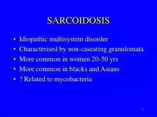
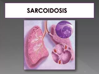
What Is It ? • Idiopathic multisystemic chronic inflammatory disease • Characterized by presence of non-caseating granulomas • Commonly affects lungs, liver, skin, eyes
Who Does It Affect ? • Worldwide – Irish and Nordic populations especially Swedes • US – African American females • Age – typically young healthy adults, unusual before 18 yoa, smaller second peak around 60 yoa • Mostly sporadic • Incidence – 20-60/100,000
Diagnosis • CXR – bilateral adenopathy • CT – more specific but not commonly used, shows peribronchial thickening and reticular nodular changes • BAL – increased lymphocytes, rule out other stuff • Mediastinoscopy • Bx – non-caseating granulomas • Supportive tests – ACE level, Ca2+ level
Signs and Symptoms • Asymptomatic to organ failure and death • Cough and SOB – most common • Non-specific skin lesions • Ocular symptoms – uveitis and optic neuritis • Constitutional – fever, night sweats, weight loss, fatigue
CXR • Initial test and most commonly used • Scoring system – Scadding in 1961 • 1 – hilar adenopathy, especially R paratracheal • 2 – adenopathy and pulmonary infiltrates • 3 – pulmonary infiltrates • 4 – fibrosis, predominantly upper lobes
Stage 1 1-2-3 sign
Stage 1 paratracheal adenopathy
Stage 2 nodules along fissures and bronchovascular bundles
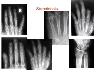
Prognosis • For most, disease is usually self-limiting and resolves within 3-5 years • Patients who present with pulmonary fibrosis, skin lesions other than erythema nodosum, bone lesions, cardiac disease, neurologic disease, or renal disease are at increased risk for chronic disease
Sources • Harrison’s online • Radiology Department of the Rijnland Hospital, Leiderdorp and the Academical Medical Centre, Amsterdam, the Netherlands • Medscape • Current Diagnosis and Treatment in Pulmonary Medicine