Download
1 / 62
620 likes | 784 Views
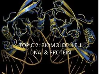
TOPIC 2: BIOMOLECULE 1 DNA & PROTEIN. Genetic information. usually. maybe. Deoxyribonucleic acid. Ribonucleic acid. Transcribed to. Held together by. Can become. Made of. Made of. Double helix. Stabilized by. Is three main. has. Linked nucleotides. Single stranded. Exist in. has.

E N D
Genetic information usually maybe Deoxyribonucleic acid Ribonucleic acid Transcribed to Held together by Can become Made of Made of Double helix Stabilized by Is three main has Linked nucleotides Single stranded Exist in has has Hydrogen bonds e.g Anti parallel strands sugar bases Phosphate ester link Denaturation/ Replication e.g bases Adenosine Cytosine Guanine Thymine Uracil rRNA tRNA mRNA Several forms Participate in Contains code for e.g Structural hierarchy translation B, A, Z Is synthesis of Protein Primary Secondary Tertiary e.g is is e.g Histones sequence 3D confirmation Supercoiling e.g Condense DNA in chromatin
DNA overview • Hallmark of life – the ability to produce • The unique information for each individual must be preserved and passed to progeny • All life on earth uses nucleic acids for storage genetic information • Except for viruses; all life use deoxyribonucleic acid (DNA) to store information
Central dogma of Molecular Biology • Sequential genetic information transferred from DNA residue to synthesis protein • DNA play essential role in heredity by serving as template for its replication. • DNA cannot flow directly to synthesis a protein • Genetic information from DNA is transferred to RNA through transcription • The sequence of RNA is translated into a protein sequence
Structure and components of the nucleotides • Nucleic acid consist of nucleotide monomer
Nucleic acids consist of repeating nucleotide that have phosphate ester, a pentose sugar, and a heterocyclic base. Phosphate Group O O=P-O O 5 CH2 O N Nitrogenous base (A, G, C, or T) C1 C4 Sugar (deoxyribose) C3 C2
Nucleoside • Base bound to pentose sugar • Pentose sugar attached to ribose – ribonucleoside • Pentose sugar attached to deoxyribose- deoxyribonucleosides
Types of nucleic acid • DNA – Deoxyribonucleic acid • RNA – ribonucleic acid
Structure and component of the nucleotides • Nucleotide also called as nucleic acid base • Base- refer to the nitrogen aromatic compound • Nucleic acid base type: pyrimidine and purine • Pyrimidine – single ring • Purine – double ring
Nitrogen-Containing Bases Uracil generally only in RNA Thymine generally only in DNA
Nucleosides in DNA Base Sugar Nucleoside Adenine (A) Deoxyribose Adenosine Guanine (G) DeoxyriboseGuanosine Cytosine (C) DeoxyriboseCytidine Thymine (T) DeoxyriboseThymidine
Nucleosides in RNA Base Sugar Nucleoside Adenine (A) ribose Adenosine Guanine (G) ribose Guanosine Cytosine (C) riboseCytidine Uracil (U) ribose Uridine
Formation of Nucleic Acid Structure • Polymerization nucleotides form nucleic acids • The phospodiester links the 5’OH of one residue and 3’OH of the next • One end must terminate at in 5’OH, the other terminates at 3’OH. Β-glycosidic bond 3 5 3’-5’ Phosphodiester bonds
Nucleic acid structure Single letter represent individual base Sequence of bases are unique and make each of us different!
Double helix structure of DNA • Determination of double helix structure was based on the X-ray diffraction patterns • Amount of T equal to A • Amount of G equal to C • Consist of 2 polynucleotide chain (we call it DNA Strand) that wrapped to each other to form helix • Chain run in antiparallel directions: • 5’ to 3’ – sense strands • 3’ to 5’ – antisense strands • Sugar phosphate backbone – outer part • Bases pair is complementary: • A—T (2 H bond) • G – C (3 H bond)
DNA Sequence Length of DNA sequence depends on organism: Bacteria e.g. Salmonella ~ 4Mb (4 million) Human ~ 3.4 Gb (billion) • kb (= kbp) = kilo base pairs = 1,000 bp • Mb = mega base pairs = 1,000,000 bp • Gb = giga base pairs = 1,000,000,000 bp.
DNA sequence • DNA sequence is obtained through Sequencing method • Sequence can be uploaded in NCBI database • Sequence of interest can also be found in the website We will find time to review this webpage later…
Denaturation of DNA • A process by which double stranded DNA unwinds and separates into single stranded strands through the breaking of hydrogen bonding between the bases • Can be achieved through heating the DNA in solution • Complete denaturation- ~94°C • Temperature needed depends on the base content of DNA; High G-C content will need a higher temperature. And why?
Renaturation of DNA Reformation of complementary strands that were separated by heat by slow cooling process
Functional role of proteins in mammalian organism • Catalysis in chemical transformations-enzymes • Transport – • Hemoglobin and myoglobin transport O2 in blood and muscle • Transferrin transport iron in blood • Metabolic control- enzymes involve in the process • Contraction – myosin and actin function in muscle contraction • Matrix for bone and connective tissue – collagen and elastin form the matrix of bone and ligament • Α-keratin- in hair and other epidermal tissue
AMINO ACID COMPOSITION OF PROTEINS • All different type of proteins are synthesized as polymers of only 20 amino acids R Group- uniquely define each of amino acid
R Groups of Amino Acids Is used to classify amino acids: • Polar or non polar • Acidic or basic
Amino acids can act as both acids and bases CO2H – Can be deprotonated to become negative carboxylates (COO-) – cause acidity NH2- – Can be protonated to become positive α- ammonium groups (+NH3) – cause basic properties
Amino acid as zwitterion Basic group Acidic group H transfer Zwitterion – a condition when amino acids are without charged groups on their side chain – no net charge; in solution- neutral
Adding alkali to amino acid solution + [OH+] Now this aa in negative charge! Donate > [H+] to bind with +[OH+] NH3+ become NH2 ( only –ve charge in COO- left)
Adding acid to acid amino solution + [H+] Now this aa in positive charge!
Amino acid charge General condition
Amino acids are polymerized into peptides and proteins • Isoelectric pH- the pH at which a molecule has no net charge – also called as isoelectric point (PI value) • The PI- allow protein to be separated using electrophoresis, isoelectric focusing and ion exchange chromatography
TASK 1: Explain the function of plasma protein in diagnosis of animal disease *Must include charge interaction and electrophoresis idea
Two shape of proteins: • Fibrous protein • Globular protein • Fibrous protein • Provide mechanical support • Often assembled into large cables or threads e.g: α-keratin – major components of hair and nails collagen – major components of tendons, skin, bones and teeth • Involved in structure :tendon, ligaments, blood clots – collagen and keratin • Contractile protein in movement: muscle, microtubule (cytoskleton, mitotic spindle, cillia, flagella)
Globular protein • Usually water soluble, compact roughly spherical • Hydrophobic interior, hydrophilic surface • Globular protein include enzyme carrier and regulatory protein • Most protein which move around (e.g albumin, casein in milk) • Proteins with binding site: • Enzymes, haemoglobin, immunoglobulin, membrane receptor sites
The peptide bond • Peptide- short polymers of amino acid monomers linked by peptide bonds • Polypeptide chain – longer peptide chain
Hierarchy structure of protein Primary structure (A.A sequence) • Secondary structure (α-helix and β-pleated sheet) • Tertiary structure (3-D structure formed by assembly of • secondary structure) • Quaternary structure (structure formed by more than • one polypeptide)
Primary structure of proteins • Sequence of amino acid in polypeptide chain • Is held together by peptide bonds • Two ends – N terminus and C terminus
Secondary structure • Local 3-D folding of the polypeptide chain in the protein • Arrangement in space of the atoms in the peptide backbone • Two type: α-helix and β-pleated sheet β-pleated sheet α-helix
Secondary structure • Forces involve: • Strong – covalent bond • Weak – hydrogen bond, electrostatic interactions, hydrophobic effect
Tertiary structure • 3D arrangement of all atoms in the proteins, including those in side chains and in prosthetic group • Describes the folding and other contortions of a polypeptide chain that result from the molecular interactions among the R groups of the different amino acids • The folding is sometimes held together by strong covalent bonds (cystein-cystein disulphide bridge) • 3-D structure is determined through X-tray crystallography • Now can be predicted using bioinformatic technique