Fatigue in Exercise Physiology: Causes and Effects
180 likes | 370 Views
This outline explores central and peripheral fatigue during exercise, identifying factors such as phosphagens, glycogen, pH levels, and potassium. It discusses the impact of environmental conditions like heat and humidity on endurance performance and how fatigue can be a result of depletion or metabolic byproduct accumulation. Additionally, it covers the exhaustion hypothesis, glycogen depletion, blood glucose levels, and the accumulation of lactic acid. The text delves into the complexities of fatigue in relation to muscle performance and cellular mechanisms, shedding light on the interplay between physiological and psychological factors.

Fatigue in Exercise Physiology: Causes and Effects
E N D
Presentation Transcript
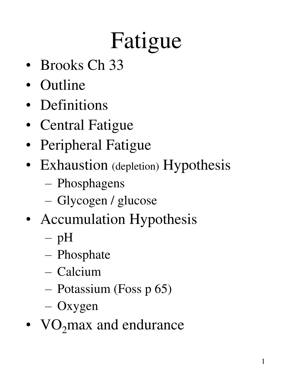
Fatigue • Brooks Ch 33 • Outline • Definitions • Central Fatigue • Peripheral Fatigue • Exhaustion (depletion) Hypothesis • Phosphagens • Glycogen / glucose • Accumulation Hypothesis • pH • Phosphate • Calcium • Potassium (Foss p 65) • Oxygen • VO2max and endurance
Fatigue During Exercise • Fatigue- inability to maintain a given exercise intensity • rarely completely fatigued - can maintain lower intensity output • Studied with EMG and observation of contractile function with electrical (nerve) or magnetic stimulation(cortex) • Observe reduction in force and velocity and a prolonged relaxation time • The effect of exercise at an absolute or relative exercise intensity will be more severe on an untrained individual • Causes of muscle fatigue have been classified into central and peripheral • Central - includes CNS, motivation and psychological factors • restoration of force with external stimulation of muscle -indicates central fatigue • NH3, hypoglycemia, reticular formation • Peripheral - PNS to muscle - EC coupling, energy supply and force generation
Identifying site of Fatigue • fatigue can be identified specifically - eg. Glycogen, Ca++ depletion • Compartmentalization within the cell increases the difficult of determining the source of fatigue • eg. ATP may be depleted at the myosin head, but adequate elsewhere in the cell - is this detectable? • Often the origin of fatigue is diffuse • eg dehydration • several factors then contribute to a disturbance of homeostasis • Often easier to identify correlations to fatigue, rather than causal contributions to fatigue
Environment and Fatigue • Heat and humidity - can affect endurance performance • inc sweat, heat gain, dehydration, changes in electrolytes results in • redistribution of Cardiac Output • Uncoupling of mitochondria - less ATP with same VO2 • changes in psychological perception of exercise • Fatigue is cumulative over time • dehydration yesterday can influence performance today • Glycogen depletion cumulative as well • Reduced circulation to muscle may result in glycogen depletion • Reducing endurance capacity
Central Fatigue • possible to have fatigue w/out the muscles itself being fatigued • eg pain may affect drive to continue • Compare force output during fatigue with force output during maximal external stimulus • An ability of this external stimulation to restore force would indicate central fatigue • Central fatigue - Stechnov Phenomenon • Fig 33-8 - faster recovery of strength with distraction or “active pauses” during recovery from exhaustion • Psychological Fatigue • understanding is minimal • With training - athletes can learn to minimize influence of sensory inputs • Able to approach performance limits
Peripheral Fatigue • Fig 33-5 - ulnar stimulation is constant - force development decrease - peripheral • Fig 33-6 - large increase in EMG signal - no increase in force - peripheral fatigue • Two hypothesis for peripheral fatigue • a) Exhaustion - depletion of energy substrates - eg ATP, CP, glycogen • Phosphagens are present in low quantities • Must match use with restoration from other metabolic pathways - or fatigue • b) Accumulation of metabolic byproducts - eg H+, NH3, Pi • Likely a combination of factors from both. Contributions of factors are influenced by the specific conditions of the activity
Exhaustion Hypothesis • Depletion of metabolites • Phosphagens • Fig 33-1a - CP levels decline in two phases - drop rapidly, then slowly • both severity of first drop and extent of final drop related to work intensity - • fig 33-2 • fatigue - in super-max cycling - coincides with CP depletion in ms • tension development related to CP level - therefore CP related to fatigue • Fig 33-1b - ATP well maintained • compartmentalization? • Down regulation / protection theory? • ms cell shuts off contraction - with ATP depletion in favor of maintaining ion concentration gradients and cell viability
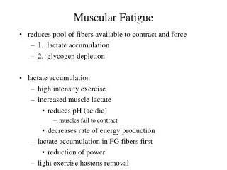
Depletion (continued) • Glycogen • depletion associated with fatigue • moderate activity - uniform depletion from different fiber types • Also activity specific fiber depletion • Carbohydrate loading can improve performance • Caffeine (inc FFA mobilization) can also offset fatigue • Blood Glucose • During short intense exercise bouts - blood glucose rises • With prolonged activity- blood glucose may fall • Anapleurotic substrates • Krebs cycle intermediates - decline results in reduced capacity of Krebs
Accumulation Hypothesis • H+ (acidity) • Lactic acid accumulates during short term high intensity exercise • As production exceeds removal • exported into blood from muscle • As it is a strong acid -blood pH decreases • H+ in blood - affects CNS • pain, nausea, discomfort, disorientation • inhibits O2 / Hb combination in lung • reduces HS lipase - dec FFA oxidation • **still unsure if this induces fatigue** • muscle acidosis • all glycolytic intermediates are weak acids • ATP breakdown also produces H+ • may inhibit PFK - slowing glycolysis • may interfere with calcium binding TnC • may stimulate pain receptors
Accumulation • Phosphate( Pi) and Diprotenated phosphate (H2PO4) • phosphagen depletion (CP) - results in Pi accumulation • behaves like proton • inhibiting PFK • interfering with X-bridge attachment • Fig 33-3 H2PO42- acid and Pi • indicative of non steady state - fatigue • Calcium Ion Accumulation • mitochondrial coupling efficiency • some Ca++ stimulates Krebs cycle • accumulation - requires energy to remove the calcium • Creates oxidative phosphorylation uncoupling in test tube • exacerbated by reduced Ca++ sequestering by SR with fatigue
Calcium (cont) • Fig 33-4 - changes in Ca++ flux and signaling in fatigued muscle • Po refers to max isometric force • symptoms of fatigue • decreased force generation - with single or tetanic stimulation • related to SR Ca++ release, and/or pH affects on opening of SR channels • 1. dec free calcium • May be EC coupling at sarcolemma, T tubules, or SR channels • Accumulation in mito, dec SR uptake • 2. Responsiveness - downward shift • H+ interference with Ca++ binding • 3. Sensitivity - small L-R shift • given free Ca++ - less force • less impact than dec release or responsiveness
Potassium (K+) • Foss p 65 • K+ is released from contracting muscle resulting in • reducing cytosolic and an increasing plasma K+ content • Release high enough to block nerve transmission in T tubules • Concomitant increase in Na+ intracellulary disrupts normal sarcolemmal membrane potential and excitability • High Na+/K+ pump activity improves performance • Rapid recovery of K+- 2-5 minutes • Complete in ~30 minutes • During exercise inactive tissues take up K+
O2 depletion and Mitochondria • O2 depletion and Mito density • dec in ms O2 or circ O2 can lead to fatigue eg - altitude, circulation impairments • low O2 often indicated by lactate accumulation, CP depletion or both • exercise depends on integration of many functions - any upset -- fatigue • Doubling of oxidative capacity with training • increases use of FFA -sparing glycogen • Minimizes impact of the damaging effects of free radicals
Heart Fatigue • Heart as site of Fatigue • no direct evidence that heart is site of fatigue • Arterial PO2 is maintained during exercise, heart gets CO priority • heart can utilize lactate or FFA • ECG - no signs of ischemia at maximal effort or fatigue • if there are signs- heart disease is indicated • With severe dehydration... Cardiac arrhythmia is possible
VO2 max and Endurance • Relationship between Max O2 consumption and upper limit for aerobic metabolism is important • Two possibilities - • 1. VO2 max limited by O2 transport • CO and Arterial content of O2 • 2. VO2 max limited by Respiratory capacity of contracting ms. • Conclude - • VO2 max set by O2 transport capacity • endurance determined by respiratory capacity of muscle • Evidence • Muscle Mass used- influences VO2max • Minimum of 50% of total ms mass for true value of VO2 max • but, at critical muscle mass VO2 max is independent of muscle mass
Muscle Mitochondria • Correlation observed between VO2 max and Mito activity - 0.8 • Henriksson - observed changes in ms mito and VO2 with Tx and detraining • ms mito inc 30%, VO2 19% • VO2 changes more persistent with detraining than respiratory capacity • illustrating independence of these factors • Davies - CH 6 - Correlation's • VO2 and End Cap .74 • Ms Resp and Running endurance.92 • Training 100% increase in ms mito • 100 % inc in running endurance • 15% inc in VO2 max • Again illustrating independence of VO2 max and endurance
VO2 and Mito • Davies study 2 - iron deficiency • Fig 33-9 restoration of iron in diet • hematocrit and VO2 max responded rapidly and in parallel • ms mito and running endurance - more slowly, but also in parallel • further experiments • anemic blood replaced with healthy blood containing red blood cells • immediately raises Hb - and restores VO2 max to 90% of pre anemic levels • running endurance was not improved • strongly suggest - VO2 max function of O2 transport • Endurance - more dependant on ms mito capacity
Future of Fatigue • Technology is making available new devices - further investigation of fatigue • NMR • possible to determine [ ] of Phosphagens, protons, water, fat, metabolites • without breaking the skin • Fig 33-10 • a at rest - before fatigue • b after fatigue • area under curve representative of [ ] of metabolites (ATP, CP, Pi) • Clear indication of declines and accumulations at fatigue • Table 33-1 comparison of values • NMR vs muscle biopsy