Download
1 / 9
90 likes | 213 Views
Presentation of the family with inherited enlarged parietal foramina. Vesna Simi ć 1 , Dejan Mirčić 2 , M omir Jovanović 1 1 General Hospital Ćuprija, Department of Neurosurgery, Miodraga Novakovića 78, 35230 Ćuprija, Serbia .

E N D
Presentation of the family with inherited enlarged parietal foramina Vesna Simić1, Dejan Mirčić2, Momir Jovanović1 1General Hospital Ćuprija, Department of Neurosurgery, Miodraga Novakovića 78, 35230 Ćuprija, Serbia. 2State University of Novi Pazar, Department of biomedical sciencies,, Vuka Karadžića bb, 36300 Novi Pazar, Serbia.
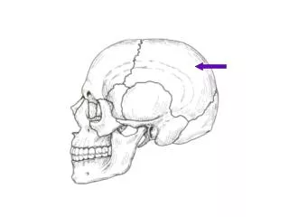
Introduction • Enlarged parietal foramina is an iherited autosomal dominant disorder which occurs during the embrionic development. • It is characterized by enlarged openings in the parietal bones, mainly symmetrical and circular with different diameters. • It is caused by deficient ossification during the first five months of the intrauterine development.
Aim The aim of our researchwas to diagnose the condition in all members of the family suffering from the disorder using the clinical diagnostic methods.
Material and methods • Roentgenography of the skull (RTG) in four live family members with enlarged parietal foramina. • Computerized tomography of the brain (CT) in four live family members with enlarged parietal foramina.
Results • Patient K.Lj. (44) admitted to the General Hospital in Ćuprija as an emergency with the head injuries after the traffic accident. The patient suffered from unconsciousness headache, nausea, slowed communication, slight disorientation, but had no neurological deficit.
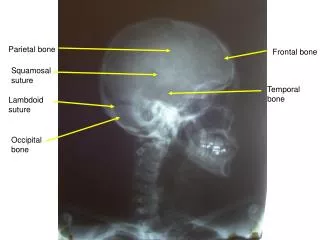
Results • During the clinical examination of the skull, the two openings in the pate region were noticed. • RTG revealed the complete skull defect, circular in shape, in the pate region. • CT also indicated the full- thickness parietal skull defects.
Results • With the insight in the family history and after the recovery of the patient, it was determined that the same disorders was found in her grandmother, mother and both sons.
Results • However, her sons’ opnenings were uneven, unlike in the female members of the family who had symmetrical dents.
Conclusion Further research will be pointed towards the usage of the molecular genetic methods in order to identify the exact gene which mutation led to the disorder in this family.