Download
1 / 15
160 likes | 358 Views
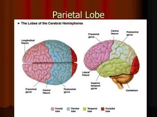
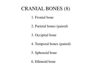
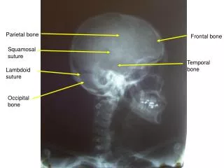
Parietal bones. Two bones, right and left, forming much of the lateral aspects of the cranium. They meet at the top of the skull at the sagittal suture . They unite with the frontal bone at the coronal suture. OCCIPITAL BONE. Thick bone forming the posterior wall and floor of the cranium.

E N D
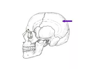
Parietal bones • Two bones, right and left, forming much of the lateral aspects of the cranium. • They meet at the top of the skull at the sagittal suture. • They unite with the frontal bone at the coronal suture.
OCCIPITAL BONE • Thick bone forming the posterior wall and floor of the cranium. • Most prominent feature is the foramen magnum, a large opening through its inferior surface.
Bordering both sides of this opening are rounded processes called occipital condyles, which articulate with the first vertebra (the atlas) for head movement.
TEMPORAL BONES • Two bones on either side of the cranium below the parietal bones. • Along the inferior margin of each temporal bone are several important features
The opening is the external auditory meatus. • Anterior to this opening is a depression called the mandibular fossa, which provides an articular (joint) surface for the mandible.
A bridgelike extension of bone that projects anteriorly is the zygomatic process, which joins the zygomatic bone to form the cheekbone, or zygomatic arch.
SPHENOID BONE • A single butterfly-shaped bone, the sphenoid is wedged between other bones in the anterior margin of the cranium.
When the bone is viewed by looking through the orbits, a round hole that penetrates the medial wall may be seen. This is the optic foramen
ETHMOID BONE • A small bone anterior to the shpenoid bone. • Portions of the ethmoid bone form sections of the cranial floor, orbital walls, and nasal-cavity walls.
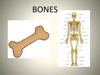
FACIAL BONES • The bones of the facial skeleton include 13 immovable bones and a movable lower jaw.
Maxillary bones • two bones on each side of the face that form the upper jaw. • Palatine bones • Zygomatic bones • Nasal bones • Lacrimal bones
Vomer • Inferior nasal conchae • Mandible
HYOID BONE • The single bone is a unique part of the skeleton, since it does not articulate with any other bone. • It is located in the neck region below the mandible, where it is suspended from the styloid process by ligaments and muscles.
The horseshoe shaped hyoid bone supports the tongue and provides attachment for some of the its muscles.
VERTEBRAL COLUMN • Vertebral columnacts as a strong, flexible rod that supports the trunk while permitting anterior, posterior, rotational, and lateral movements.