THE NERVOUS SYSTEM
520 likes | 539 Views
This article provides an overview of the divisions of the nervous system, neuroglial cells, neuron anatomy, classification of neurons, nerve physiology, and the role of neurotransmitters. It explains the concepts in a detailed yet accessible manner.

THE NERVOUS SYSTEM
E N D
Presentation Transcript
Divisions of the NS • Central Nervous System (CNS) • Peripheral Nervous System (PNS) Fig. 11.32
Sensory vs. Motor Nerves • SENSORY nerves: • Body CNS • MOTOR nerves: • CNS Body Fig. 10.2
Somatic NS consciously controlled effectors Autonomic NS involuntary effectors 2 Different Types of Motor Nerves
NEUROGLIAL CELLS NEURONS vs. Cells of the Nervous System
NEUROGLIAL CELLS • Fill spaces • Provide structure • Produce myelin • Phagocytize bacteria & cellular debris • Outnumber neurons • Can divide (mitosis)
Neuron Anatomy Overview • Dendrites Cell body Axon Synaptic knobs at axon terminals Effector
Neuron Anatomy • Schwann cells • type of neuroglial cell • myelin sheath • Nodes of Ranvier Fig. 10.3
THE DIRECTION THEIR SHAPE Classification of Neurons Fig. 10.6
Sensory, Motor, and Interneurons (Direction) • Sensory neurons • PNS CNS • Motor neurons • CNS PNS • Interneurons • in between sensory and motor neurons Fig. 10.7
Shapes of Neurons Fig. 10.6
Neuron vs. a “Nerve” • Neuron = a cell • Nerve = bundles of neuron axons, and neuroglial cells bound together • outside brain/spinal cord Fig. 11.24
Neuron Physiology • Sending neuron impulses = action potential • change in electrical charge in cell membrane • depends on electrolytes • potassium (K+) and sodium (Na+)
First Things First: Creating a Resting Potential • Protein pumps • open and close • let ions through • Active pumps • against a gradient • Passive pumps • with the gradient Fig. 10.13 Na+/K+ Pump
Resting Potential Fig. 10.14
Action Potential Fig. 10.15 Action Potential
A Nerve Impulse- a series of action potentials Fig. 10.16 Computer activity http://outreach.mcb.harvard.edu/animations/actionpotential.swf
Action Potential Fig. 10.18 Action Potential Zoomed Out
What happens when the nerve impulse reaches the end of the axon? • axon terminals • next to another neuron (as shown) or a muscle or gland • Gap called a synapse Synapse Fig. 10.11
The Synapse • Neurotransmitters • Synaptic cleft • Receptors • Send a message Fig. 10.12
EXCITATORY = depolarize the next neuron It tells the next neuron/muscle/gland to GO INHIBITORY = hyperpolarize the next neuron prevent the nerve impulse from continuing It tells the next neuron/muscle/gland to STOP Classification of Neurotransmitters
First neurotransmitter discovered (1921) Mostly excitatory Skeletal muscle neuromuscular junctions & synapses between the brain and spinal cord Message = muscles contract or continue sending impulses Acetylcholine (ACH)
Acetylcholine cont. • Nicotine • Activates acetylcholine receptors • Releases dopamine (coming later…) • Alzheimers • Memory loss, depression, disorientation, dementia, hallucinations,death • Deficient acetylcholine
Generally excitatory helps send messages in the brain Involved in learning and memory Alcohol inhibits glutamate receptor function Monosodium Glutamate (MSG) food additive stimulates glutamate receptors in the taste buds Glutamate
Serotonin • Found in the brain • Primarily inhibitory • Sleep, mood and temperature regulation • Insomnia – deficient serotonin • Antidepressants (Prozac, Zoloft, Paxil, etc) • “SSRI’s” or Selective Serotonin Reuptake Inhibitors • Serotonin accumulates in the synapse • feel more content • LSD blocks serotonin • MDMA releases excess serotonin
Dopamine • AKA “the brain reward” • Regulates emotions, moods and subconscious control of skeletal muscle • Nicotine • excess dopamine release • Cocaine • blocks reuptake (leaves more in the synapse) • Methamphetamine • excess dopamine release
Dopamine - cont’d • Dopamine also sends signals that help coordinate your skeletal muscle movements • Parkinson’s Disease • deficient dopamine production • tremors
Found in the brain Generally inhibitory Prevents the receptor nerve from being overstimulated When it accumulates it has a sedative effect Valium, Xanax and Ativan work by allowing GABA to accumulate Huntington’s Disease – deficient GABA GABA
Found in the brain Alertness, regulation of moods Ritalin & Adderall- increase level of norepi and dopamine Strattera- increase only norepi Clinical depression – low norepi Norepinephrine
Endorphins • Flood the synaptic cleft during pain or stress • Usually inhibit neurons from firing, causing an analgesic effect • At lower levels can excite the next neuron • Reduces pain and makes one feel good • “Opiates” (heroin, codeine, morphine, oxycodone, hydrocodone, etc) • bind to endorphin receptors and mimic endorphins
Anandamide • Involved in working memory, regulation of feeding behavior, generation of motivation and pleasure • Anandamide receptors are called cannabinoid receptors • A lot of cannabinoid receptors in the hippocampus (short term memory), cerebellum (coordination) and basal ganglia (unconcious muscle movement) of brain • THC (found in marijuana) mimics anandamides and binds to cannabinoid receptors
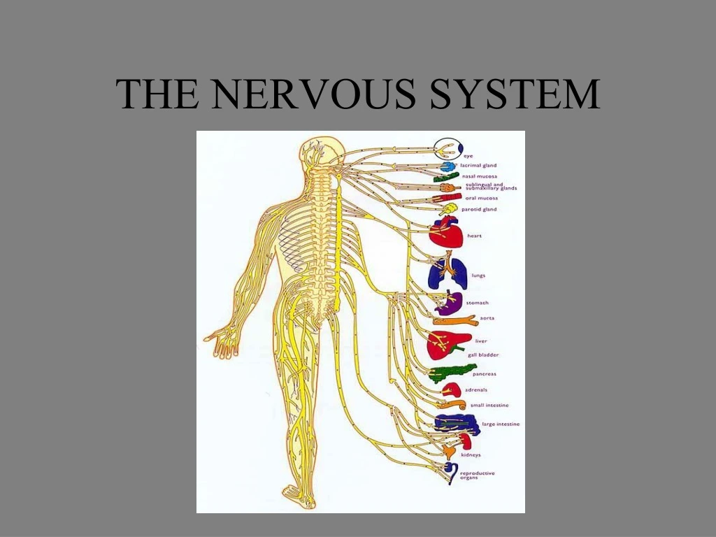
12 pairs cranial nerves 31 pairs spinal nerves Peripheral Nervous System
PNS Flow Chart Peripheral Nervous System Motor Sensory nerves nerves Somatic Autonomic nerves nerves Sympathetic Parasympathetic nerves nerves
PNS cont. • Motor nerves are divided into • Somatic n.s.- conscious activities • Autonomic n.s. – unconscious activities • Autonomic n.s is divided into • sympathetic and • parasympathetic divisions.
SYMPATHETIC “fight or flight” responses speeds up heart rate, breathing and other functions vital to survival Digestion and other less essential functions will be slowed for awhile. PARASYMPATHETIC when the body is not mobilized and active in fight or flight. speeds up digestion and other essential functions When the body is in this mode, heart rate and breathing are calm. PNS cont.
The Brain The Spinal Cord The Central Nervous System
Central Nervous System - Spinal Cord Figs11.5, 11.6, & 11.7
Reflexes Fig. 11.8
Central Nervous System: The Brain • Cerebrum • Largest part • Sensory & motor functions • Higher mental functions (memory, reasoning, etc) • Brainstem • Connects the cerebrum to the spinal cord • Cerebellum • Coordinates voluntary muscle movements • Diencephalon • Processes sensory info Fig. 11.15
The Cerebrum • Divided into right and left cerebral hemispheres • Covered by folds called convolutions/gyri and grooves called sulci (little groves) and fissures (big grooves) • Connected by the corpus callosum • It has a cortex: an outer covering about 2 mm thick • Gray matter vs. white matter
The Cerebrum cont. • The cerebral cortex is divided into LOBES which control various functions • FPOT Fig. 11.16 & 11.17
FRONTAL LOBE – “Primary Motor Area” controls voluntary muscles “Broca’s Area” motor speech usually L hemisphere Voluntary eye movement Concentration, planning, problem solving, analysis The Cerebrum cont.
The Cerebrum cont. • PARIETAL LOBE • Sensory info: touch, taste, pressure, pain • interpretation of sensory info, “awareness” of body • “Wernicke’s Area” • sensory speech, understanding written & spoken language • usually L hemisphere
The Cerebrum cont. • OCCIPITAL LOBE • visual senses • analyzing visual patterns, combining visual images with other info (i.e. recognizing a person) • TEMPORAL LOBE • sensory smell and hearing • interpretation of sensory experiences (understanding speech, reading)
Cerebral Hemispheres • Hemisphere = half of sphere (brain) • The right side of the brain controls the left side of the body and vice versa • Corpus callosum
The Cerebellum • Integrates sensory info • Balance, coordination of skeletal muscle, posture
Brainstem • Brainstem: Connects the cerebrum to the spinal cord • Midbrain: visual and auditory reflex center • Pons: transfer nerve impulses • Medulla Oblongata: • Cardiac center- heart rate • Vasomotor center- smooth muscle in blood vessels/blood pressure • Respiratory center- breathing rate • Coughing, sneezing, swallowing and vomiting reflexes Fig. 11.21
Diencephalon • Thalamus- - Receives all sensory impulses (except smell) and relays them to the appropriate region of the cerebral cortex 2. Hypothalamus – • Maintain homeostasis • Links the nervous system to the endocrine system 3. Pituitary & pineal glands Fig. 11.19
Diencephalon cont. • The limbic system is a collection of structures involved in emotional behavior and your feelings • Includes the amygdala and hippocampus


