Download
1 / 17
210 likes | 1.08k Views
SPLANCHNOLOGY . Chapter 10 The Peritoneum. General description Relationship between the abdominopelvic viscera and peritoneum Structures formed by the peritoneum Subdivision of the peritoneal cavity. General description.

E N D
SPLANCHNOLOGY Chapter 10 The Peritoneum General description Relationship between the abdominopelvic viscera and peritoneum Structures formed by the peritoneum Subdivision of the peritoneal cavity
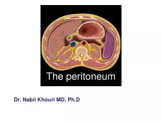
General description The peritoneum lines the wall of abdominal and pelvic cavities, and covers the abdominal and pelvic viscera. It is composed of a single layer of mesothelium supported by a thin layer of connective tissue termed the serous membrane parietal peritoneum The peritoneum lining the wall of abdominal and pelvic cavities is parietal peritoneum。 visceral peritoneum covering the viscera peritoneal cavity The potential space between the parietal and visceral peritonea is the peritoneal cavity
Relationship between the abdominopelvic viscera and peritoneum Intraperitoneal organs almost completely surrounds with peritoneum the stomach, superior part of duodenum, jejunum, cecum, vermiform appendix, transverse colon, sigmoid colon, spleen, ovary, uterine tube Interperitoneal organs covered by peritoneum on their three aspects the liver, gallbladder, urinary bladder, uterus, upper part of the rectum, ascending and descending colon. Retroperitoneal organs Only the anterior part of these organs is covered by the peritoneum the kidneys, suprarenal glands, ureters, major part of duodenum, pancreas (except for its tail), and lower part of the rectum
Structures formed by the peritoneum OmentaThe omentum is double-layered extension or fold of peritoneum that connects the greater curvature or lesser curvature of the stomach Lesser omentum • hepatogastric ligament • hepatoduodenal ligament • right anterior :the bile duct • left anterior : the proper hepatic artery • posterior:hepatic portal vein
Greater omentum • gastrocolic ligament ⑴
Omental bursa and omental foramen omental bursa (lesser peritoneal cavity ) There is a potential, narrow space between the lesser omentum, the posterior wall of the stomach and the posterior wall of the abdomen anterior wall the lesser omentum, the posterior wall of the stomach and the gastrocolic ligament posterior wall the peritoneum covering the pancreas, the left kidney and suprarenal gland superior wall the inferior surface of the caudate lobe of the liver and the peritoneum covering diaphragm inferior wallthe transverse mesocolon and the site that the anterior and posterior layers of the greater omentum fuse the left the spleen, the gastrosplenic and splenorenal ligament . the right the omental bursa communicates with the greater sac through the omental foramen.
omental foramen (foramen of Winslow) is located posterior to the hepatoduodenal ligament (free edge of lesser omentum), upper boundThe caudate lobe of the liver lower boundThe superior part of the duodenum prozonethe hepatoduodenal ligament Post boundThe inferior vena cava
Mesenteries mesentery mesoappendix transverse mesocolon sigmoid mesocolon
Ligaments Ligaments of the liver falciform ligament ligamentun teres hepatis coronary ligament right and left triangular ligaments
Ligaments of the spleen gastrosplenic ligament splenorenal ligament phrenicosplenic ligament
Ligaments of the stomach Hepatogastric ligament gastrosplenic ligament gastrocolic ligament gastrophrenic ligament phrenicocolic ligament
Folds, peritoneal recesses, and pouches Folds and peritoneal recesses of posterior abdominal wall the superior duodenal fold superior and inferior duodenal recesses retrocecal recesses intersigmoid recess
Folds and peritoneal fossae of anterior abdominal wall median umbilical fold ⑴ medial umbilical folds ⑵ lateral umbilical folds ⑶ supravesical fossa⑷ medial inguinal fossa(superficial inguinal ring ) ⑸ lateral inguinal fossa (deep inguinal ring) ⑹ rectus abdominis ⑴ parietal peritoneum ⑶ ⑵ ⑹ ⑸ ⑷ urinary bladder
Pouches Male:rectovesical pouch ⑴ Female: the rectouterine pouch (Douglas pouch) ⑵ the vesicouterine pouch ⑶ ⑵ ⑴ ⑶
Subdivision of the peritoneal cavity subphrenic space The peritoneal cavity is divided into the supracolic and infracolic compartments by the transverse colon and transverse mesocolon Suprahepatic space left anterior suprahepatic spaces left posterior suprahepatic spaces right anterior suprahepatic spaces right posterior suprahepatic spaces left suprahepatic spaces right suprahepatic spaces
Subhepatic space left subhepatic spaces left anterior and left posterior subhepatic spaces right subhepatic spaces
Paracolic sulci Mesenteric sinuses Infracolic compartment left paracolic sulcus right paracolic sulcus right mesenteric sinus left mesenteric sinus