Download
1 / 22
420 likes | 3.43k Views
The anterolateral abdominal wall and peritoneum . Zahir Mughal SCRUBS. Introduction. By the end you will take away with you the following: Boundaries of the abdominal cavity Layers of the abdominal wall Inguinal canal Inguinal hernias But you won’t learn much about:

E N D
The anterolateral abdominal wall and peritoneum Zahir Mughal SCRUBS
Introduction • By the end you will take away with you the following: • Boundaries of the abdominal cavity • Layers of the abdominal wall • Inguinal canal • Inguinal hernias • But you won’t learn much about: • Peritoneum and peritoneal cavity
Inguinal Canal “Questions on the anatomy of this region are probably asked more often than any other in examinations because of its importance in diagnosis and treatment of hernias” Harold Ellis
Abdominal cavity Anterior boundary Anterolateral abdominal wall Upper boundary Thoracic Diaphragm Posterior boundary Posterior abdominal wall Lower boundary Imaginary plane at the level of the pelvic brim
Regions Midclavicular point Midclavicular point Subcostal plane Transumbilical plane Interstubercular plane Midinguinalpoint Midinguinal point Median plane
Quadrants • Liver (left lobe) • Spleen • Stomach • Jejunum and proximal ileum • Pancreas (body and tail) • Left kidney • Left adrenal gland • Left colic flexure • Transverse colon (left half) • Descending colon (superior part) • Liver (right lobe) • Gallbladder • Stomach (pylorus) • Duodenum (parts 1-3) • Head of the pancreas • Right adrenal gland • Right kidney • Right colic flexure • Ascending colon (superior part) • Transverse colon (right half) • Sigmoid colon • Descending colon (inferior part) • Left ovary • Left uterine tube • Left ureter (abdominal part) • Left spermatic cord • Uterus (enlarged) • Urinary bladder (full) • Cecum • Appendix • Most of ileum • Ascending colon (inferior part) • Right ovary • Right uterine tube • Right ureter (abdominal part) • Right spermatic cord • Uterus (enlarged) • Urinary baldder (full)
QUIZ 1 • What organs in the abdominal cavity are protected by the: 1. rib cage? • Spleen, Liver, Stomach, part of kidneys 2. Pelvic girdle? • Cecum, sigmoid colon, ilium
Quiz • Two causes of acute abdominal pain in: • 1. Right upper quadrant • Biliary disease. Peptic ulcer disease • 2. Right lower quadrant • Appendicitis. Ectopic pregnancy • 3. Left upper quadrant • Pancreatitis. Peptic ulcer disease • 4. Left lower quadrant • Diverticulitis. Ectopic pregnancy • 5. Diffuse or central • Gastroenteritis. AAA • 6. Flank • Peylonephritis. Pneumonia
Understanding the Anterolateral Abdominal Wall • Musculoaponeurotic wall is made up of several layers: • Skin • Camper fascia • Scarpa fascia • External oblique • Internal oblique • Transversusabdominis • Transversalis fascia • Extraperitoneal fat • Parietal peritoneum
Abdominal Muscles • Five muscles: (bilaterally paired) • External oblique • Internal oblique • Transversus abdominis • Rectus abdominis • Pyramidalis
Aponeuroses of external oblique, internal oblique and transversusabdominis form the rectus sheath
Quiz • 1. What is the name of the fibrous structure that runs down the midline of the abdomen? • Linea alba • 2. What is the name of the three fibrous structures thatrun horizontally? • Transverse tendinous intersections • 3. What two layers of the abdominal wall do the neurovascular structures run between? • Internal oblique and transversusabdominis • 4. Challenging question - What is the name of the line that marks the point where both layers of the rectus sheath move anterior to the rectus muscles. • Arcuate line of Douglas
Functions of the abdominal muscles • Increase intra-abdominal pressure • Forced expiration • Bladder emptying • Vomiting • Excretion of faeces (and flatus) • Childbirth (expulsion of fetus) • Protect the abdominal viscera
Understanding Inguinal Canal • Need to know a bit about the embryology • Inguinal canal represents an oblique passage taken by the testis and spermatic cord (round ligament in females)
Inguinal Canal • Downwards and medially • 4 cm long • Parallel and superior to inguinal ligament
Boundaries • Anteriorly • External oblique aponeurosis • Internal oblique lateral 1/3 • Posteriorly • Medially – Conjoint tendon • Laterally – Transversalis fascia • Roof • Internal oblique and transversusabdominis • Floor • Inguinal ligament • Two ends • Superficial ring / external ring • Deep ring / internal ring
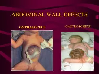
Hernias • Abnormal protrusion of a viscus • Through the wall of the cavity that normally contains it. • Enclosed in a hernial sac.
How to clinically diagnose inguinal hernias? • Ask yourself FOUR questions: • 1. Inspection of the location of the swelling • Inguinal or inguino-scrotal or scrotal? • 2. Palpate over superficial ring. Is there a cough impulse? • 3. Is the hernia reducible? • 4. Is it direct or indirect? Palpate the deep ring with your hand while the hernia is reduced and ask the patient to cough. • Hernia will not appear if it is indirect • Hernia will appear if it is direct
Peritoneum • A serous membrane – two layers • Parietal layer • Visceral layer • Thin layer of peritoneal fluid between the two • Function • Allow movement of the abdominal viscera • Transmit nerves, lymphatics and blood supply from the abdominal wall to the viscera • Mesenteries are double-layered peritoneum that transmit neurovascular structures