1 / 108

CXR in Respiratory Distress

CXR in Respiratory Distress. John T. Lysack, MSc, MD, FRCPC Clinical Associate Professor of Radiology, Clinical Neurosciences, and Surgery, Faculty of Medicine, University of Calgary. Normal Chest X-Ray (CXR) Anatomy & Pathology Lungs Pleura Heart Bones. Overview. Normal CXR.

Download Presentation

CXR in Respiratory Distress

An Image/Link below is provided (as is) to download presentation Download Policy: Content on the Website is provided to you AS IS for your information and personal use and may not be sold / licensed / shared on other websites without getting consent from its author. Content is provided to you AS IS for your information and personal use only. Download presentation by click this link. While downloading, if for some reason you are not able to download a presentation, the publisher may have deleted the file from their server. During download, if you can't get a presentation, the file might be deleted by the publisher.

E N D

Presentation Transcript


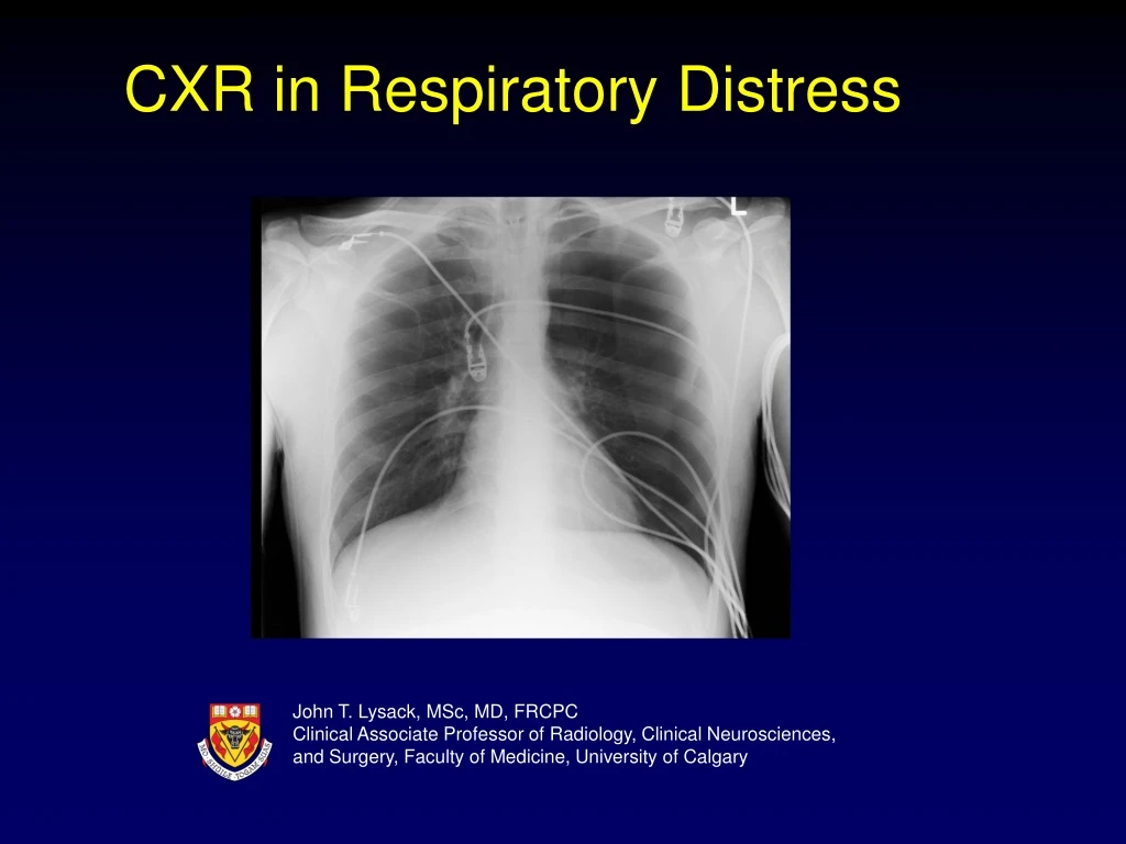
  1. CXR in Respiratory Distress John T. Lysack, MSc, MD, FRCPC Clinical Associate Professor of Radiology, Clinical Neurosciences, and Surgery, Faculty of Medicine, University of Calgary

  2. Normal Chest X-Ray (CXR) Anatomy & Pathology Lungs Pleura Heart Bones Overview

  3. Normal CXR

  4. Anatomy – Cardiac Right Atrium

  5. Anatomy – Cardiac Right Ventricle

  6. Anatomy – Cardiac Left Atrium Left Atrium

  7. Anatomy – Cardiac Left Ventricle Left Ventricle

  8. Anatomy – Cardiac Aortic Arch Aortic Arch

  9. Anatomy – Cardiac Pulmonary Arteries

  10. Anatomy – Lung – Right Upper Lobe Minor Fissure Minor Fissure

  11. Anatomy – Lung – Right Middle Lobe Minor Fissure Minor Fissure Major Fissure

  12. Anatomy – Lung – Right Lower Lobe Major Fissure

  13. Anatomy – Lung – Left Upper Lobe Major Fissure

  14. Anatomy – Lung – Left Lower Lobe Major Fissure

  15. Anatomy – Pleura

  16. Anatomy – Bones Ribs Clavicle Ribs Spine Spine

  17. Normal CXR

  18. Pathology – Lung

  19. Atelectasis vs. Consolidation • Atelectasis • Volume loss • Adjacent segments hyperinflate • Mucus plugging, splinting • Rapidly changes on X-ray • Consolidation • No volume loss • Air bronchograms • Pneumonia, edema, hemorrhage • Slowly changes on X-ray

  20. Atelectasis – RUL

  21. Atelectasis – RUL

  22. Atelectasis – RML

  23. Atelectasis – RML

  24. Atelectasis – RUL & RML

  25. Atelectasis – RUL & RML

  26. Atelectasis – LUL

  27. Atelectasis – LUL

  28. Bronchograms

  29. Consolidation – Air Bronchograms

  30. Consolidation – Air Bronchograms

  31. Pneumonia – RUL

  32. Pneumonia – RUL

  33. Pneumonia – RML

  34. Pneumonia – RML

  35. Pneumonia – RLL

  36. Pneumonia – RLL

  37. Pneumonia – LUL

  38. Pneumonia – LUL

  39. Lung Abscess

  40. Lung Abscess Fluid Level Fluid Level

  41. Lung Abscess

  42. Tuberculosis

  43. Tuberculosis

  44. Tuberculosis

  45. Tuberculosis

  46. Lung Cancer

  47. Lung Cancer This particular case looks identical to a lung abscess Fluid Level

  48. Lung Cancer

  49. Lung Cancer This case has the more typical appearance of a tumor

  50. Lung Metastases

More Related