Download
1 / 1
Download Presentation 
caso1jfa_copy1
An Image/Link below is provided (as is) to download presentation
Download Policy: Content on the Website is provided to you AS IS for your information and personal use and may not be sold / licensed / shared on other websites without getting consent from its author.
Content is provided to you AS IS for your information and personal use only.
Download presentation by click this link.
While downloading, if for some reason you are not able to download a presentation, the publisher may have deleted the file from their server.
During download, if you can't get a presentation, the file might be deleted by the publisher.
E N D
Presentation Transcript
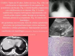
CASO1: Varón de 45 años, fiebre sin foco. Fig. 1:Rx con calcificación en hipocondrio D con aspecto de granuloma calcificado. Fig. 2: lesión hepática hipoecoica con calcificación en su interior. Fig. 3: Lesión multilocular irregular heterogénea hipoatenuada con calcificación central, catéter drenaje en su interior.Derrame pleural D acompañante. Fig. 4:Correlación histológicadel granuloma necrotizante: halo de células epitelioides mezclada con polimorfonucleares, restos nucleares, linfocitos y células plasmáticas. Internamente necrosis caseiforme con macrófagos (publicada en Diagnostic Microbiology and Infectious Disease. (2002) Mar ; 42 159-167)..
More Related