
Download
1 / 1
Download Presentation 
copy1
An Image/Link below is provided (as is) to download presentation
Download Policy: Content on the Website is provided to you AS IS for your information and personal use and may not be sold / licensed / shared on other websites without getting consent from its author.
Content is provided to you AS IS for your information and personal use only.
Download presentation by click this link.
While downloading, if for some reason you are not able to download a presentation, the publisher may have deleted the file from their server.
During download, if you can't get a presentation, the file might be deleted by the publisher.
E N D
Presentation Transcript
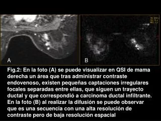
A B Fig.2: En la foto (A) se puede visualizar en QSI de mama derecha un área que tras administrar contraste endovenoso, existen pequeñas captaciones irregulares focales separadas entre ellas, que siguen un trayecto ductal y que correspondió a carcinoma ductal infiltrante. En la foto (B) al realizar la difusión se puede observar que es una secuencia con una alta resolución de contraste pero de baja resolución espacial

More Related