Download
1 / 17
190 likes | 565 Views
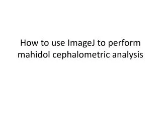
How to use ImageJ to perform mahidol cephalometric analysis. Step one. Download the two excel files Download ImageJ and install it on your computer http://rsbweb.nih.gov/ij/download.html. Step two. Open the software Click the File button, the Open

E N D
Step one • Download the two excel files • Download ImageJ and install it on your computer • http://rsbweb.nih.gov/ij/download.html
Step two • Open the software • Click the File button, the Open • Open the x-ray you want to analyze. You may need to convert it to jpg first
Step 3 • The Basic command of imageJ: moving • Use the scrolling tool • Or press space bare and left click on the mouse
Step 3 • The Basic command of imageJ: Zoom • Zoom in + • Zoom out - • Or go in : image > transform > zoom
Step 3 • The Basic command of imageJ: brightness and contrast • Go to Image > Adjust > Brightness/contrast • A window will appear, Keep it on the side of the work space
Step 4 • Open the excel file for male or female depending on the patient gender and put in on the side of the screen to know in what order you have to select the point • You should have the main window of imageJ and the little window for the contrast
Step 5 • Click each point is respecting the sequence • You can move each point just by clicking on it • An example with all the point
Step 5 • For the point 31 an 32 you have to indicate the real distance in mm between the point • For the knew X ray machine the distance between the point is 31 mm • For the old machine just use the ruler on the side : in this example you have to put 35 mm in the case 33 • For the PA analysis there is no ruler you have to measure a distance on the film manually and use it as a reference on the scan
Step 6 Saving your point • When finish go to File> Save as • Save as > selection that will create a .roi file ( this will allow you to keep your point ) • Save as > XY coordinate this will create a .txt file ( to use with excel )
Step 7 • Open the .txt file the coordinate of the point are display • Copy everything • Select the case of the X coordinate of point S • Past
Step 7 The coordinate will be transfer to excel
Step 8 • Enter the real distance you use as reference on point 33 • Magnification : the norm of Mahidol analysis have been calculated with a magnification of 7% • So by default this value is 1.07 if you want to use Mahidol norm for a Thai patient DON’T TOUCH IT • If your patient is not Thai ask your Professor for advice • This is important only for measurement in MM for the angle it does not make any difference
Step 8 • THE EXCEL SHEET • This column give you the variation to the norm in percentage • 100 to 25% the patient is normal • 25 to 0 % the patient is Tendency ( Tendency prognathic, retrognatic, open , close configuration …) • The value is negative the patient is outside the norm, the more important the value the more the patient is outside the norm
Step 8 • This column give you the variation to the norm in Degree and millimeter • The value is positive the patient is inside the norm • The value is negative the patient is outside the norm and you know by how many degree or millimeter • For the value in millimeter there is automatic correction of the magnification, in this example the patient mandible length is 3.37 mm to longue.
Step 9 • The PA You do exactly the same IF YOU SEE ANY MISTAKE PLEASE LET ME KNOW
Step 10 MOTIVATE TOTO TO ADD AUTOMATIC COLORING DEPENDING ON THE RESULTS