Download
1 / 16
170 likes | 328 Views
Quantifying Glial and Axonal Damage due to Blast Injury in Rat Model. Dr. Kurosh Darvish & Dr. Servio Ramirez Temple University Presented by Christine Yoo. Inspiration for Research. Armed conflicts with Afghanistan and Iraq

E N D
Quantifying Glial and Axonal Damage due to Blast Injury in Rat Model Dr. KuroshDarvish & Dr. Servio Ramirez Temple University Presented by Christine Yoo
Inspiration for Research • Armed conflicts with Afghanistan and Iraq • “One report noted that 88% of Operation Iraqi Freedom(2003)-associated injuries treated at a second-echelon military medical center were the result of blasts” [IPPITO] • >1700 soldiers have sustained a TBI since 2006 [IPPITO] • Issues with Injury • Explosions and blasts • High survival rate • Long-term effects
Injury Due to Blast Wave 1 2 3 4
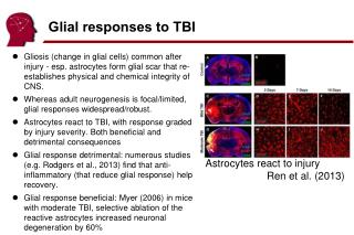
Previous Research • Exploratory research to understand the mechanisms behind how the brain reacts due to blast overpressure (BOP) • Damage can occur directly or indirectly to the brain - Dr. IboljaCernak • Measured rupture pressure: 183 ± 14 kPa • BBB permeability increases (marked by IgG marker) • 3 days to recover
Goals • To establish brain injury severity as it relates to shockwave intensity • To evaluate the progression of injury in the brain following blast induced trauma
Hypothesis • The relationship between how the shock wave intensity and brain damage will occur exponentially and then plateau predictably enough to create a referrable chart to diagnose a patient
Controls • Mice controls • C57BL/6 strain • Approximately 7 weeks • Male • Cranial window installed vs. cranial window absent • Pressure sensor implanted vs. pressure sensor absent • Calibration of shock tube
Variables • Amount of time for damage to spread or heal • Variation of blast wave overpressure intensities • Position of the mouse • Location of the blast wave on the mouse’s head or body
Methodology • Na-Fl permeability in BBB • Histology • transcardial perfusion, brain removal, blood collection and histology • First anesthetized • Then series of experimentations • Then respective methods of review
Potential Problems • Genone – different enzymes and processes – healing processes • Different shape of skull presents problem with introducting blast wave damage to frontal area
Discussion/Implications • Create better preventative measures • Can prepare medical doctors to better understand the level of damage due to BOP as well as move forward in the healing process
Reference • Bolger, Frank, Ben Sauers, and Ray Cornely. Design and Fabrication of a Shock Tube For Conducting Brain Injury Experiments in Rats. Senior Design. Temple University. PowerPoint. • Cernak, Ibolja. "The Importance of Systemic Response in the Pathobiology of Blast-Induced Neurotrauma." Frontiers in Neurology 1.151 (2010): 1-9. Print. • Ippolito, Charles J. "Battlefield TBI: Blast and Aftermath." Psychiatric Times 3.8 (2007). UBM Medica LLC, 1 Aug. 2007. Web. 17 June 2011. <http://www.psychiatrictimes.com/display/article/10168/56916>. • Readnower, Ryan D., MikulasChavko, and SaleenaAdeeb. "Increase in Blood–brain Barrier Permeability, Oxidative Stress, and Activated Microglia in a Rat Model of Blast-induced Traumatic Brain Injury." Journal of Neuroscience Research 88.16 (2010): 3530-539. Print.
http://www.nature.com/nprot/journal/v4/n8/images/nprot.2009.89-F1.jpg - cranial window • http://www.mc.uky.edu/coa/images/neuron%20cartoon.jpg – microglial activation cartoon • http://www.utwente.nl/ewi/bios/research/Cellsonchips/barrier.doc/barrier-1.jpg - BBB description • http://www.tacticalwarfightergear.com/graphics/2010prototype.jpg - soldier • http://images.the-scientist.com/content/figures/images/yr1998/oct/oct_art/3d_rats.jpg - mouse skel