Download
1 / 47
470 likes | 565 Views
Join us in BIOL 260 to explore the world of microbes! Learn about prokaryotes, eukaryotes, historical developments, and microscopic techniques. Understand the role of microorganisms in health and disease. Discover the fascinating realm of microbiology!

E N D
BIOL 260-General Microbiology Instructor: Christopher Thor Masters Degree, Bioengineering Bachelors Degree, Molecular Biology
Welcome to BIOL 260: Microbiology! • First day: • Review of Syllabus • Sign-in • Introduce the course, review course expectations • Begin with first lab • Exercise 3: Microscope Lab
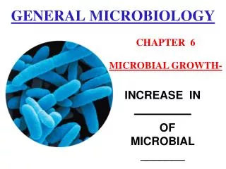
Objectives for today • Define prokaryotes, eukaryotes & their classification • Give a historical perspective on medical bacteriology • Introduction to bacterial stains and images
What is microbiology? • The scientific discipline which studies microbes or microorganisms • Biology of microbes • The interaction of microbes with other microbes, the environment, and humans
The “Yotes” Definitions: Prokaryote: Single celled organism, no nucleus. Bacteria, Archaea Eukaryote: Single or multi-celled organism, membrane bound nucleus Algae, Protozoa, Fungi, people
What are examples of microbes? • Algae • Fungi • Protozoa • Bacteria • Viruses Which are Prokaryotes are which are Eukaryotes?
Microorganisms are associated with • Disease • Cause of many epidemics in history • Bubonic plague (1346-1350) • Killed 25 million people • Small pox • Killed estimated 600 million people since 10,000 BC • Eradicated in 1979 • HIV • 3.1 million estimated new cases per year • 5% of Sub-Saharan Population • Malaria Small Pox
Bacteria are associated with • Normal microbiota (normal flora) • The bacteria that are present on our bodies
Bacteria are associated with • The environment • Rhizobium (the greatest bacteria you’ve never heard of) • Nitrogen fixation in the soil • Food products • Beer! Or bread, wine, sauerkraut, yogurt, cheese… • Medicines • Bacteria are “programmed” to make insulin
It all started with the microscope! Zacharis Janssen (1600) Antoni van Leewenhoek (1632-1723) Robert Hooke (1665) History of Microbiology
Zacharis Janssen’s microscope • Modeled after the telescope • Consisted of two lenses • Magnified images 3-10X
Leewenhoek’s microscope 20-30x magnification
Where do cells come from? • Spontaneous generation • Francesco Redi (1668) • Spontaneous Generation does not occur • John Needham (1745) • Spontaneous Generation does occur • Lazzaro Spallanzani (1765) • Louis Pasteur (1861) • Biogenesis • Rudolf Virchow (1858) • Living things come from living things
John Tyndall questions Pasteur’s experiments • Could not reproduce Pasteur’s results • Specific growth media required • Found that there were heat resistant forms of microbes • Same year (1876) Ferdinand Cohn discovers heat resistant forms of bacteria called endospores • Spores can survive in space (Apollo Program, 1960s) • 1877 Robert Koch demonstrates that anthrax caused by Bacillus anthracis
New cells need to be placed in categories • Aristotle-plant or animal kingdom • Kingdom Protista (1866) • Electron microscope (1940’s) • Kingdom Procaryotae (1968) • Carl Woese proposed 3 Domains (1978)
Prokaryotes (Single Celled) • Bacteria Domain (Eubacteria) • Peptidoglycan cell walls • Gram negative • Gram positive • Archaea Domain (Archaebacteria) • Not a peptidoglycan cell wall • Extremophiles • Methanogens • Halophiles • Thermophiles
Binomial system of nomenclature • Genus and species • Escherichia coli, Escherichiacoli • E. coli is not acceptable on exams or unknowns… • Both names are in italics or underlined and correctly spelled.
Types of microscopes • Brightfield • Darkfield • Phase Contrast • Fluorescent • Electron
Stained specimen Microscopy, Oil Immersion Wet mount
Microscopy Electron microscopes - maximum magnification 100,000X
Microscopy Electron microscopes - maximum magnification 100,000X “Color-enhanced”
Relative sizes Figure: CNX.org
Staining: key to visualization • Simple • Differential • Special
Microscopic Techniques: Dyes and Staining • Simple stains • Stains everything • Differential stains • Stain based on cellular traits Gram stain - separates bacteria into two categories based on type of cell wall Acid Fast Stain – Stains non-peptidoglycan containing bacteria (Mycobacteria) Gram-positive Gram-negative
Microscopic Techniques: Dyes and Staining • Simple stains • Differential stains Gram stain - separates bacteria into two categories based on type of cell wall Purple: Bacteria with high peptidoglycan containing cell walls Pink: Counter stain
Microscopic Techniques: Dyes and Staining Fluorescent dyes and tags
Morphology of Prokaryotic Cells: Multicellular Associations Biofilm containing mixed species